At birth, the vitreous is in a gel-like state. Around the age of 4, initial signs of vitreous liquefaction begin to appear. Vitreous liquefaction refers to the gradual dehydration and shrinkage of the gel-like vitreous, leading to a separation of water and collagen. By the ages of 14 to 18, approximately 20% of the vitreous cavity is comprised of liquid. Between the ages of 45 and 50, the water content within the vitreous increases significantly, while the gel-like components decrease. By the ages of 80 to 90, more than 50% of the vitreous is liquefied. In elderly individuals, further liquefaction of the vitreous leads to vitreous detachment. The separation of the vitreous from the posterior lens capsule is referred to as anterior vitreous detachment, while separation from the internal limiting membrane of the retina is known as posterior vitreous detachment (PVD). The incidence of PVD is approximately 58% in individuals over 50 years of age and 65% to 75% in those over 65 years of age.
Histopathological Changes
With advancing age, histological changes in the vitreous include:
- Gradual depletion and dissolution of hyaluronic acid result in the destabilization of collagen. Portions of the collagen network collapse, forming liquid pockets that are surrounded by collagen fibers, a process referred to as vitreous dehydration and contraction (syneresis).
- Vitreoschisis, characterized by splitting within the vitreous cortex.
- Liquefied vitreous material enters the posterior vitreous cavity through cortical fissures. Initially, partial separation between the vitreous and retina occurs, ultimately progressing to complete posterior vitreous detachment.
- Thickening of the basement layer (internal limiting membrane of the retina), accompanied by loosening of attachments to the posterior retina.
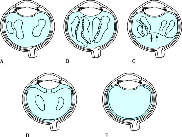
Figure 1 Age-related changes in the vitreous
A: Formation of vitreous liquefaction cavities
B: Liquefaction and collagen fiber appearance
C: Posterior vitreous detachment
D: Anterior vitreous detachment
E: Thickening of the basement layer
In addition to age, other conditions such as aphakia, intraocular inflammation, vitreous hemorrhage, and elongated axial length may also lead to PVD.
To be continued