Anatomical Overview
A distal radius fracture refers to a fracture within 3 cm of the distal articular surface of the radius. This region is a transition zone between cancellous (trabecular) bone and cortical bone, making it anatomically weaker and prone to fracture when subjected to external forces. The distal articular surface of the radius is concave, sloping from the dorsal to the palmar side and from the radial to the ulnar side, forming the volar tilt (10°–15°) and ulnar tilt (20°–25°). The distal ulnar side of the radius articulates with the radial side of the ulnar head to form the distal radioulnar joint, which, along with the proximal radioulnar joint, provides the anatomical basis for forearm rotation. The radial styloid process is located 1–1.5 cm distal to the ulnar styloid process. Together, the distal ends of the radius and ulna articulate with the proximal carpal bones to form the wrist joint.
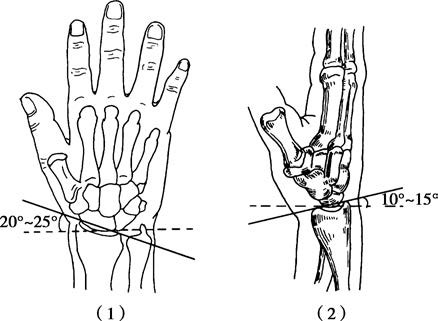
Figure 1 Normal ulnar and volar tilt of the radiocarpal joint
(1) Ulnar tilt
(2) Volar tilt
Etiology and Classification
Most distal radius fractures are caused by indirect trauma. When falling, if the hand is outstretched and the ground impact force is transmitted upward, it can result in a distal radius fracture. Depending on the mechanism of injury, different types of fractures can occur, including extension-type fractures, flexion-type fractures, and articular fractures with wrist dislocation.
To be continued