Among abdominal arterial aneurysms, splenic artery aneurysms rank second only to infrarenal abdominal aortic aneurysms and iliac artery aneurysms, making them the most common type of visceral arterial aneurysm. Splenic artery aneurysms are commonly located in the distal one-third of the splenic artery and near the splenic hilum. They are usually solitary and exhibit a saccular or spherical dilation.
Etiology
Splenic artery aneurysm development is associated with the following factors or conditions:
Pregnancy
It is more frequently observed in pregnant women and carries a high risk of rupture, with rupture rates as high as 20% to 50%.
Portal Hypertension
Portal hypertension can lead to splenomegaly and increased blood flow within the splenic artery, resulting in aneurysmal dilation of weaker segments of the arterial wall.
Pancreatitis
Both acute and chronic pancreatitis can trigger the formation of pseudoaneurysms in the splenic artery due to autodigestion by pancreatic enzymes or localized pressure.
Trauma
Major abdominal surgeries may directly damage the splenic artery, leading to the formation of pseudoaneurysms. Intraluminal vascular interventions that cause direct damage to the arterial wall are another contributing factor to aneurysm formation.
Clinical Manifestations
The clinical presentation of splenic artery aneurysms varies widely. Symptoms are often nonspecific in unruptured cases, with some patients experiencing only upper abdominal discomfort or pain. Larger aneurysms may cause pain in the left shoulder or back. Compression of the celiac plexus or irritation of the posterior gastric wall may result in intermittent gastrointestinal symptoms such as nausea or vomiting. Ruptured aneurysms typically lead to sudden onset of acute abdominal pain, radiating pain in the back or shoulder, and signs of acute hypovolemic shock.
Diagnosis
CTA
This imaging modality can accurately distinguish the splenic artery and the aneurysmal dilation. Three-dimensional reconstructions allow visualization of the aneurysm’s spatial structure from various angles.
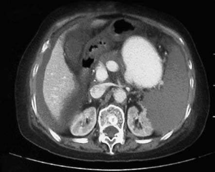
Figure 1 CTA imaging of splenic artery aneurysm
MRI
Magnetic resonance imaging can utilize its vascular flow void effect to assist in diagnosing splenic artery aneurysms, while additionally assessing blood flow within the portal vein and visceral veins.
Ultrasound
Although its diagnostic sensitivity is lower than that of CTA and MRI, it serves as a preliminary screening tool.
Selective Angiography
Most commonly performed as digital subtraction angiography (DSA), this method provides detailed information about the aneurysm’s size, shape, location, and relationship to surrounding structures. It is also useful for planning endovascular treatment.
Treatment
Treatment is generally indicated for aneurysms with a diameter of ≥2 cm, aneurysms showing a tendency to enlarge, or aneurysms discovered during pregnancy or in women planning to become pregnant. Two main approaches are available for treatment:
Endovascular Therapy
This minimally invasive approach includes techniques such as aneurysm embolization or placement of covered stents to exclude the aneurysm.
Open Surgery
Options include aneurysm resection, splenic artery reconstruction, or concurrent splenic artery aneurysm resection and splenectomy.