Spinal arteriovenous malformation (AVM) is a rare condition, occurring more frequently in males than females, with 80% of cases presenting between the ages of 20 and 40. Spinal AVMs develop slowly and may remain stable for many years. These malformations can be located intramedullary and/or extramedullary and may also form arteriovenous fistulas outside the dura mater. Given the varying blood supply to different regions of the spinal cord, spinal AVMs are anatomically classified into three segments: cervical, upper thoracic, and lower thoracic-lumbar-sacral. The latter is the most common.
Clinical Manifestations
Compression of the spinal cord or nerve roots caused by the AVM may result in segmental symptoms, including numbness and reduced muscle strength in the affected limbs.
Rupture of malformed vessels can lead to subarachnoid hemorrhage or intramedullary hematoma. Patients often present with acute pain corresponding to the spinal cord level of the malformation, with pain that may be triggered by changes in body position. Clinical symptoms also commonly include intermittent claudication, limb weakness or paralysis, and sphincter dysfunction.
Imaging Characteristics
MRI scans typically show flow voids indicative of vascular malformations. In some cases, an abnormal thread-like signal of intermediate intensity can be observed on T2-weighted images. In cases with associated hemorrhage, the lesion may include irregular, punctate regions of high intensity on short T1 inversion recovery (STIR) sequences. MRI is also helpful in distinguishing spinal AVMs from intramedullary cavernous malformations (CM). Spinal angiography is effective in determining the location and extent of the AVM.
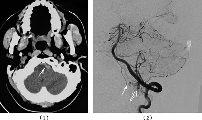
Figure 2 Subarachnoid hemorrhage, associated with spinal anterior artery aneurysm and vascular malformation
(1) CT image showing a small subarachnoid hemorrhage on the left anterior side of the medulla oblongata (arrow).
(2) Spinal anterior artery aneurysm (solid arrow), associated with vascular malformation (hollow arrow), as shown on imaging.
Treatment
Surgical resection by microsurgery is highly effective for superficial, localized spinal AVMs. For more extensive spinal AVMs, endovascular treatment may be utilized.