Traumatic intracerebral hematoma is relatively uncommon, with an incidence of approximately 0.5% to 1.0% in closed head injuries. It is often associated with contrecoup cerebral contusions and lacerations in the frontal and temporal lobes caused by impact to the occipital region. In a small number of cases, the hematoma is located at the site of impact.
Mechanism of Occurrence
Intracerebral hematomas can be classified into two types. Superficial hematomas are mostly caused by ruptures of blood vessels in contused or lacerated cortical tissue and are often accompanied by subdural hematomas, commonly located in the frontal pole, temporal pole, and their respective bases. Deep hematomas, on the other hand, result from ruptures of deep brain vessels. Contusions and lacerations may also be present on the brain surface.
Clinical Manifestations and Diagnosis
The symptoms of intracerebral hematoma are very similar to those of subdural hematoma accompanied by cerebral contusions and lacerations. CT imaging shows round or irregular hyperdense lesions near the site of brain contusions or within the deep white matter.
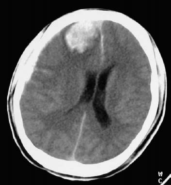
Figure 1 Intracerebral hematoma (CT, right frontal lobe)
Treatment
The treatment approach for intracerebral hematoma is similar to that for subdural hematoma. Craniotomy using a bone flap or burr hole method is usually performed to remove the intracerebral hematoma, subdural hematoma, and contused brain tissue. In cases of small, deep-seated hematomas without significant intracranial pressure elevation, surgery may not be necessary. In such cases, an intracranial pressure monitor is utilized, and the patient’s condition is closely observed.