Anemia
Anemia refers to a syndrome where the peripheral red blood cell volume is reduced below the lower limit of the normal range, resulting in insufficient oxygen transport to tissues. Ocular manifestations of anemia may include visual impairment, eye strain, or visual field defects. The severity of fundus changes depends on the type and degree of anemia, the onset speed, and individual responses. Mild anemia may present with a normal fundus appearance, but if hemoglobin levels or red blood cell counts fall to 30%–50% of normal, fundus changes can occur. The most common sign is retinal hemorrhages, frequently flame-shaped or punctate, although linear or irregular hemorrhages may also be observed, typically located in the posterior pole. Retinal vessels may appear pale; arterial diameters are often normal or slightly narrowed, while veins may become tortuous, dilated, and pale. Cotton-wool spots may be present on the retina, with occasional hard exudates. Retinal edema may be localized to the posterior pole or involve the entire retina. Severe anemia, such as pernicious anemia, can result in ischemic optic neuropathy, optic neuritis, or optic atrophy, potentially causing blindness. Sickle cell anemia may lead to proliferative retinopathy and associated findings, including conjunctival pallor, subconjunctival hemorrhages, oculomotor disturbances, nystagmus, and sluggish pupillary responses.
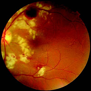
Figure 1 Anemic retinopathy
Leukemia
Leukemia refers to a group of malignant clonal diseases of hematopoietic stem cells and is characterized by symptoms such as fever, infections, bleeding, anemia, hepatosplenomegaly, and systemic organ damage. Ocular abnormalities are often associated and may result in visual impairment, vision loss, visual field defects, night blindness, or proptosis. Specific findings include:
Fundus Changes
Tortuous, dilated retinal veins, flame-shaped or deep dot hemorrhages, and sometimes preretinal hemorrhages may occur, with Roth spots being a classic finding. Retinal exudates are less common, but nodular retinal infiltrates are observed in cases with high leukocyte counts or immature leukocytes, indicating a poor prognosis. In chronic leukemia, microaneurysms may be noted in peripheral retina, occasionally accompanied by peripheral vascular occlusion and neovascularization. In acute leukemia, optic disc edema with hemorrhages and optic neuropathy may occur.
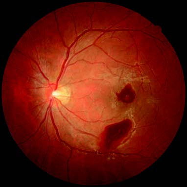
Figure 2 Fundus changes in leukemia
Orbital Infiltration
Orbital infiltration is common in young children. Acute leukemia may cause leukemic cell infiltration of orbital tissues, resulting in proptosis, restricted ocular motility, ptosis, conjunctival congestion, and edema. In some cases, especially in acute myeloid leukemia, firm masses in the orbital margin known as "chloromas" or granulocytic sarcomas may be palpable, leading to proptosis, diplopia, or blindness. Orbital infiltration suggests severe disease with a poor prognosis.
Anterior Segment Changes
These changes are most common in acute lymphoblastic leukemia. Clinical features include spontaneous subconjunctival hemorrhages, anterior chamber bleeding, pseudohypopyon, iris infiltration and thickening, and signs resembling acute iridocyclitis.
Polycythemia Vera
Polycythemia vera is a chronic myeloproliferative disorder primarily characterized by clonal overproduction of red blood cells. Peripheral blood hematocrit increases, resulting in elevated blood viscosity, often accompanied by leukocytosis, thrombocytosis, splenomegaly, thrombosis, and bleeding complications. Ocular manifestations range from normal vision to transient blurred vision, night vision disturbances, visual field defects, photopsia, floaters, photophobia, eye strain, and diplopia. Retinal vein occlusion may occur, with tortuous and dilated retinal veins appearing purplish-red or purple-black. Retinal arteries may show dilation, and retinal hemorrhages and exudates are less common but are typically confined to the superficial layers. Eyelid skin may appear purplish-red, with dilated and engorged conjunctival vessels and small hemorrhages. Expansion of superficial scleral and iris vessels may also be observed.