Kidney diseases that lead to ocular complications primarily include glomerulonephritis and chronic renal failure.
Acute glomerulonephritis is characterized by eyelid edema, often accompanied by fundus changes caused by hypertension, including retinal vascular spasm, retinal hemorrhages, and exudates. These changes are reversible and typically resolve as the underlying disease improves.
More than 50% of patients with chronic glomerulonephritis exhibit fundus changes, with approximately 75% of those experiencing renal insufficiency showing ocular manifestations. Almost all patients with uremia develop fundus alterations. These manifestations include hypertensive retinopathy and anemic retinopathy, such as narrowing of the retinal arteries, arteriovenous crossing changes, and tortuous and dilated retinal veins. Other findings may include diffuse grayish-white retinal edema, hard exudates, retinal hemorrhages, cotton-wool spots, and optic disc hyperemia and edema. These changes may gradually improve as systemic conditions stabilize. However, the prognosis is poor, and the appearance of optic disc edema and cotton-wool spots indicates an even worse prognosis.
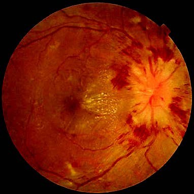
Figure 1 Retinal changes in glomerulonephritis
Chronic renal failure may also lead to band keratopathy and cataract formation. Retinal edema is often pronounced in patients undergoing dialysis. Kidney transplant recipients, due to the usage of corticosteroids and other immunosuppressants, may develop cataracts and cytomegalovirus (CMV) infection syndrome.