Arteriosclerotic Retinopathy
The characteristic features of arteriosclerosis include non-inflammatory, degenerative, and proliferative changes in the arteries, broadly classified as senile arteriosclerosis, atherosclerosis, and arteriolosclerosis. Senile arteriosclerosis primarily occurs in individuals aged 50 to 60 years and above, characterized by systemic diffuse hyalinosis and fibrotic degeneration of the arterial media. Atherosclerosis predominantly affects large and medium arteries, with the aorta, coronary arteries, and cerebral arteries being the most commonly involved. In the eye, it often involves the intra-optic nerve segment of the central retinal artery, the lamina cribrosa, and the major branch arteries near the optic disc. Arteriolosclerosis represents a reactive change caused by gradual and sustained elevation of blood pressure and is frequently associated with hypertension.
Retinal arteriosclerosis observed during fundus examination includes senile arteriosclerosis and arteriolosclerosis. These changes are considered a reflection of cerebrovascular and systemic vascular conditions to some extent and are collectively referred to as arteriosclerotic retinopathy. The primary manifestations include:
- Diffuse narrowing of retinal arteries, increased tortuosity, reduced coloration, widened arterial reflex, and straighter vessel trajectories.
- Venous concealment and arteriovenous nicking at arteriovenous crossing points.
- Exudates and hemorrhages, particularly in the posterior pole of the retina, which typically occur without edema.
Hypertensive Retinopathy
Hypertension is a cardiovascular syndrome marked by elevated systemic arterial pressure and is categorized into primary and secondary hypertension.
Primary Hypertension
Primary hypertension accounts for over 95% of all hypertension cases, with fundus changes observed in approximately 70% of patients. These changes are influenced by factors such as age, the degree of blood pressure elevation, and the duration of the condition. Older age and longer disease progression are associated with a higher incidence of fundus alterations. Retinal arteries respond to hypertension by undergoing vasospasm, narrowing, and thickening of the vascular walls. Severe cases exhibit exudates, hemorrhages, and cotton-wool spots. Fundus changes are clinically graded using the Keith-Wagener classification:
- Grade I: Characterized by arterial constriction and narrowing, with generalized thinning of the retinal arteries, and an increased arterial light reflex.
- Grade II: Arterial narrowing with arteriovenous crossing changes (arteriovenous nicking).
- Grade III: Fundus changes include hemorrhages and cotton-wool-like exudates in addition to the above alterations.
- Grade IV: Accompanies the aforementioned changes with optic disc edema.
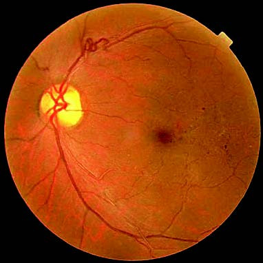
Figure 1 Hypertensive retinopathy
Acute Hypertension
Hypertensive emergencies occur when sudden and significant rises in blood pressure (generally above 180/120 mmHg) lead to progressive dysfunction in critical target organs such as the heart, brain, and kidneys. The most notable ocular manifestations include optic disc edema, retinal hemorrhages, and exudates.
Secondary Hypertension
Secondary hypertension refers to elevated blood pressure caused by identifiable conditions or factors, accounting for approximately 5% of all hypertension cases. Fundus changes in secondary hypertension may resemble those observed in primary hypertension.
In addition to developing hypertensive retinopathy, hypertensive patients may experience retinal vein occlusion, ischemic optic neuropathy, ocular motor nerve palsy, retinal artery occlusion, and exudative retinal detachment.