Eyelid Contusions and Lacerations
Contusions can cause rupture of small eyelid blood vessels, leading to edema and hemorrhage. Bruising initially appears blue-purple, gradually changing to yellow and usually being fully reabsorbed within 1 to 2 weeks. Severe contusions or sharp injuries can result in full-thickness lacerations of the eyelid, extending to the musculature, tarsal plate, or conjunctiva.
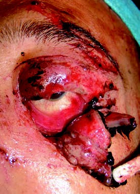
Figure 1 Full-thickness eyelid laceration
The image shows a full-thickness laceration of the left lower eyelid, along with multiple skin lacerations involving the brow and upper eyelid.
Treatment
Significant ecchymosis and swelling may be alleviated with cold compresses within the first 48 hours after the injury, followed by warm compresses later.
Eyelid lacerations require prompt debridement and suturing. Tissue preservation is essential, and skin excision should be avoided to ensure proper restoration of both functionality and cosmetic appearance. Full-thickness lacerations should be meticulously sutured layer by layer to minimize scar formation and eyelid deformities. Levator aponeurosis repair is essential in cases involving levator muscle rupture to prevent ptosis.
Eyelid repair should adhere to the following principles: The rich vascular supply of the eyelid makes ischemic necrosis rare. While partial-thickness lacerations not involving the eyelid margin may be simply sutured, other injuries require precise alignment of the eyelid margin, tarsal plate, and skin. Mattress sutures on the tarsal plate near the eyelid margin are often employed to prevent angular deformation. Early surgery is preferred since edema after 24 hours post-injury increases the complexity of suturing.
Canalicular Injury
Medial eyelid trauma often involves damage to the lacrimal apparatus, with lower canalicular injury being most common. Such injuries can result from sharp objects causing direct laceration or from sudden lateral traction on the eyelid causing indirect tearing of the delicate medial canthal structures. Improper treatment may lead to eyelid deformities and epiphora.
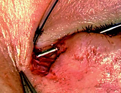
Figure 2 Canalicular laceration
The image displays a full-thickness laceration of the left lower eyelid accompanied by lower canalicular injury. During surgical repair, a lacrimal stent is inserted from the nasal end of the lacerated canaliculus, passing through the lacrimal sac and nasolacrimal duct into the nasal cavity. The temporal end of the canaliculus is identified using a metallic probe for successful alignment.
Treatment
Surgery is the only effective treatment. Early canalicular repair is recommended, ideally within 48 hours of injury. Delayed treatment can be complicated by tissue edema, which interferes with canalicular repair or reconstruction. Precise identification of the canalicular ends during surgery is critical for success and is best performed under sedation or anesthesia using a surgical microscope with good illumination. A thorough understanding of medial canthal anatomy is essential. Techniques such as using a probe or fluorescein sodium dye for irrigation can assist with localization. Under microscopic guidance, stents (e.g., epidural catheters, specialized silicone tubes, or other supports) are inserted to realign the canaliculus and surrounding tissue to their normal anatomical positions. Sutures are used to repair the canalicular wall, adjacent muscle, soft tissue, and any accompanying eyelid skin lacerations. The stent is typically removed 3 to 6 months postoperatively. If significant irritation, infection, local inflammation, or pyogenic granuloma formation develops, earlier removal of the stent is advised. In cases of combined upper and lower canalicular injury, attempts should be made to repair and anastomose both canaliculi.