This condition arises from vascular damage to the ciliary body, retina, or choroid. Minimal bleeding tends to remain localized initially but may later disperse and be reabsorbed on its own. In cases of significant hemorrhage, optical media become opaque, preventing observation of the fundus. B-scan ultrasonography is required in such instances, revealing densely packed punctate, clumped, or linear medium-to-low echoes in the vitreous cavity. Additionally, this evaluation can help determine the presence of retinal or choroidal detachment as well as posterior vitreous detachment.
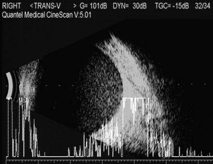
Figure 1 Vitreous hemorrhage
B-scan ultrasonography shows uniformly consistent, fine punctate medium-to-low echoes within the vitreous cavity.
In cases involving macular injury, choroidal rupture, or retinal detachment, visual recovery is typically impaired, necessitating surgical intervention.