Orbital schwannoma, also known as schwannoma, is a tumor caused by abnormal proliferation of Schwann cells that cover nerve axons. It can affect the motor, sensory, sympathetic, parasympathetic nerves, and the ciliary ganglion within the orbit. The optic nerve sheath, which lacks Schwann cells, generally does not develop schwannomas.
Clinical Manifestations
Ocular symptoms depend on the size and location of the tumor. Early stages are generally asymptomatic. Significant tumor growth may result in pronounced proptosis, globe displacement, restricted ocular motility, and decreased vision. Tumors located at the orbital apex may cause visual field defects, optic disc edema, and vision loss, even when small, and can extend intracranially through the superior orbital fissure. CT and MRI imaging often show a well-demarcated tumor. On T1-weighted imaging (T1WI), the lesion typically appears isointense, while on T2-weighted imaging (T2WI), it shows mixed intermediate-to-high signal intensity. After contrast enhancement, the tumor typically demonstrates inhomogeneous enhancement, with possible involvement extending towards the orbital apex or intracranial region.
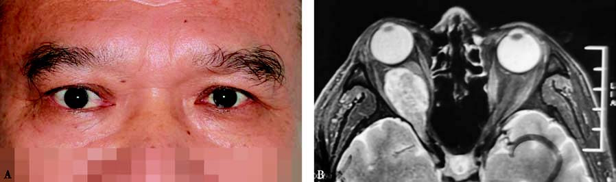
Figure 1 External appearance and imaging of a patient with orbital schwannoma in the right eye
A. External appearance shows proptosis of the right eye.
B. Axial T1-weighted MRI with contrast enhancement shows inhomogeneous enhancement of the tumor with invasion into the superior orbital fissure.
Treatment
When the lesion does not cause clinical symptoms, close monitoring is advisable. If visual function is impaired or significant proptosis occurs, surgical excision may be performed.