Uveal metastasis refers to secondary malignancy in the uveal tract originating from systemic cancers, most commonly breast cancer and lung cancer. Other primary tumors that may metastasize to the uvea include renal cell carcinoma, gastrointestinal tumors, thyroid carcinoma, hepatocellular carcinoma, and melanoma. Among uveal sites, the choroid is the most common location for intraocular metastases (88%), followed by the iris (9%) and the ciliary body (2%). The condition may involve one or both eyes.
Clinical Manifestations
Blurred vision or visual impairment is the most frequent symptom associated with choroidal metastasis. Due to the rapid growth of metastatic lesions, severe eye pain and headache may occur early if the ciliary nerves are compressed. Other symptoms include diplopia, photopsia, floaters, and visual field defects. Most choroidal metastases are located posterior to the equator and commonly appear as yellow-white or gray-brown, flat, nodular elevations in the posterior pole of the retina, often accompanied by subretinal fluid. Extensive retinal detachment may develop in advanced stages. Iris metastases demonstrate a variety of clinical presentations, including one or more yellow, white, or pink focal nodules in the iris stroma. If cancer cells disseminate into the aqueous humor, intraocular inflammation or pseudo-hypopyon may occur. Ciliary body metastases are often inconspicuous, presenting as solitary masses or mimicking features of iridocyclitis. A detailed medical history of prior malignancies, comprehensive systemic evaluation, and identification of the primary tumor are critical during diagnosis.
Diagnosis
Key diagnostic evaluations include slit-lamp biomicroscopy, anterior segment and fundus photography, ocular ultrasonography, fluorescein angiography (FFA), indocyanine green angiography (ICGA), optical coherence tomography (OCT), and magnetic resonance imaging (MRI). Biopsy can aid in determining the nature of the tumor and identifying the primary malignancy.
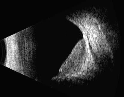
Figure 1 Ocular B-scan ultrasound of choroidal metastasis in the right eye
The image shows a broad-based flat elevation arising from the choroid, with the surface appearing slightly undulated. The internal echogenicity of the lesion is relatively homogeneous, exhibiting medium to low reflectivity.
Treatment
Systemic therapy targeting the primary malignancy is the most important treatment approach. If intraocular metastases progress and cause significant eye pain or other symptoms during systemic treatment, or if the eye is the primary metastatic site, local ocular treatment may be considered. Local treatment options include radiation therapy, cryotherapy, laser therapy, and surgical interventions.