Choroidal hemangioma is a congenital hamartoma resulting from vascular developmental abnormalities and can be classified into two types: circumscribed and diffuse. Circumscribed choroidal hemangioma presents as a solitary, orange-red spherical elevation, whereas diffuse choroidal hemangioma manifests as widespread, flat thickening of the choroid with indistinct margins. The diffuse form often occurs alongside other systemic vascular malformations, most commonly seen in Sturge-Weber syndrome.
Clinical Manifestations
Choroidal hemangiomas typically occur in young adults and are often located in the posterior pole of the eye. They may present as solitary lesions, appearing as an orange-red, round or nearly spherical elevation, or as diffuse lesions, characterized by a widespread, flat, tomato-colored thickening with ill-defined borders. These lesions can lead to exudative retinal detachment, resulting in visual impairment, and may also cause refractory glaucoma and eventual blindness.
Diagnosis
Fundus examination, optical coherence tomography (OCT), fluorescein angiography (FFA), indocyanine green angiography (ICGA), and ocular ultrasonography provide significant diagnostic value. On FFA, early stages show prominent hyperfluorescence of intratumoral blood vessels, with diffuse staining visible in later stages. ICGA can directly visualize the tumor's feeder vessels, showing early fluorescence filling of the tumor and characteristic rapid clearing of fluorescence in the late phase, referred to as the "washout phenomenon."
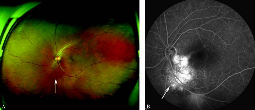
Figure 1 Wide-field fundus photograph and FFA of left eye with choroidal hemangioma
A. The wide-field fundus photograph shows a solitary, oval orange-red elevated lesion located inferior to the optic disc in the temporal region.
B. The FFA image demonstrates prominent hyperfluorescence of the lesion in the late phase of angiography.
Treatment
Asymptomatic, non-progressive choroidal hemangiomas can be monitored through regular follow-ups. Symptomatic or active choroidal hemangiomas can be treated using photodynamic therapy, transpupillary thermotherapy, radiation therapy, or anti-angiogenic therapies. Surgery may be considered when necessary.