Adenoma of the non-pigmented ciliary epithelium and adenoma of the pigmented ciliary epithelium originate from the non-pigmented and pigmented epithelial layers of the ciliary body, respectively, and are considered benign tumors. These tumors are more commonly seen in adults. Adenoma of the non-pigmented ciliary epithelium, lacking pigment, typically appears white or grayish-white in color. In contrast, adenoma of the pigmented ciliary epithelium usually presents as a gray-brown to black mass, which can easily be misdiagnosed as melanoma. The tumor's appearance, transillumination testing, and imaging examinations such as UBM and MRI are useful in distinguishing between ciliary epithelial adenoma and melanoma, although histopathological analysis remains the "gold standard" for confirming the origin and nature of the tumor.
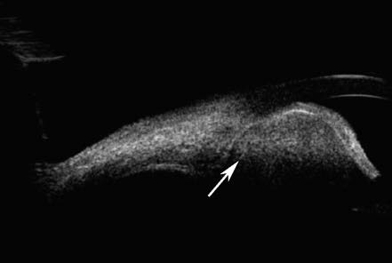
Figure 1 UBM Imaging of a non-pigmented ciliary epithelium adenoma in the left eye
The adenoma of the non-pigmented ciliary epithelium appears as a mass with inhomogeneous medium echogenicity and clear borders (white arrow).
The primary treatment approaches include follow-up observation and local surgical resection, yielding good visual and overall prognoses.