Papilloma of the lacrimal sac is the most common benign tumor of this region and often develops following chronic inflammation of the lacrimal drainage system or mucosal injury. Based on growth patterns, it can be classified into exophytic, inverted, and mixed types. Inverted papilloma, which demonstrates an aggressive nature, has a high recurrence rate and the potential for malignant transformation.
Clinical Presentation
The condition typically arises between the ages of 30 and 40 and is primarily characterized by epiphora or a painless mass located in the medial canthal region. It is usually unilateral, with the tumor exhibiting slow, expansive growth. CT dacryocystography reveals a well-defined solid mass in the lacrimal sac region with an intact capsule. The tumor may compress surrounding tissues, and prolonged pressure can lead to partial bone resorption; however, no bone destruction is observed. MRI findings show intermediate to low signals on T1-weighted images and intermediate to high signals on T2-weighted images, with significant enhancement of the lacrimal sac lesion following contrast administration. Pathologically, inverted papillomas are characterized by non-keratinized stratified squamous epithelium growing into the underlying stroma, forming areas of invasive acanthosis. Exophytic papillomas typically present as papillary or cauliflower-like projections connected to normal tissue via a narrow stalk.
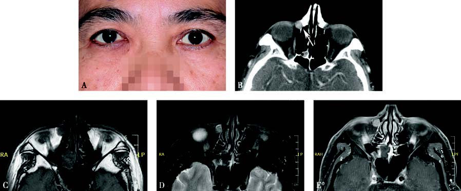
Figure 1 Appearance and imaging of a case with right lacrimal sac papilloma
A. A painless mass in the right medial canthal region of the patient.
B. Axial CT showing a solid mass in the lacrimal sac region with clear boundaries and partial bone resorption, but without invasive destruction (white arrow).
C. Axial T1-weighted MRI showing an intermediate to low signal from the tumor.
D. Axial T2-weighted MRI showing an intermediate to high signal from the tumor.
E. Contrast-enhanced axial T1-weighted MRI showing significant enhancement of the lacrimal sac lesion.
Treatment
Surgical treatment is the main approach. During surgery, the tumor is excised by opening the lacrimal sac. In severe cases, the affected nasolacrimal duct is also removed.