Lymphoma of the lacrimal gland, most commonly extranodal marginal zone lymphoma of mucosa-associated lymphoid tissue (MALT lymphoma), is a low-grade malignancy with an indolent course and a relatively favorable prognosis.
Clinical Presentation
This condition is more common in middle-aged and elderly individuals and may involve one or both sides. Typical features include proptosis with downward displacement of the eyeball and an S-shaped appearance of the upper eyelid. CT scans demonstrate diffuse enlargement of the lacrimal gland with well-defined margins and rarely show bone destruction. MRI features include intermediate to low signals on T1-weighted images and intermediate to high signals on T2-weighted images, with contrast-enhanced imaging showing significant enhancement of the lacrimal gland lesion. Pathologically, the tumor is homogeneous, soft, and friable, with a fish-flesh appearance on the cut surface. Microscopically, the lesion consists of small to medium-sized B cells.
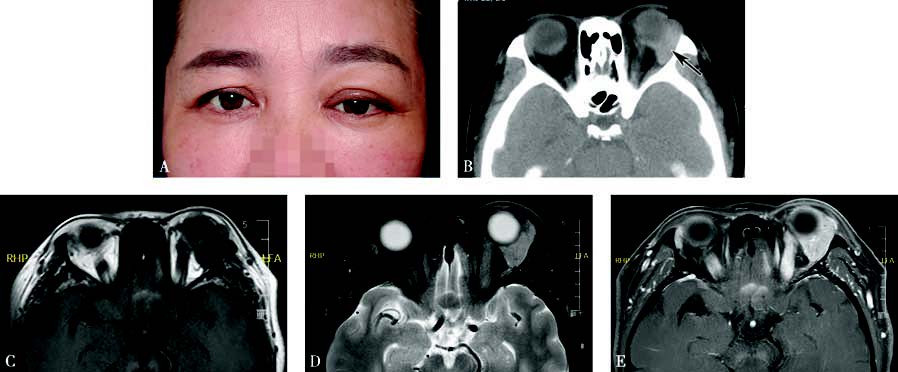
Figure 1 Appearance and imaging of lacrimal gland lymphoma
A. A photograph of a patient with lymphoma of the left lacrimal gland, showing swelling in the left lacrimal gland region, proptosis, and ptosis of the upper eyelid.
B. Axial CT imaging shows diffuse enlargement of the lacrimal gland with well-defined margins and no evident bone destruction (black arrow).
C. MRI T1-weighted imaging reveals intermediate to low signals from the lesion.
D. MRI T2-weighted imaging with fat suppression shows intermediate signals from the tumor.
E. Contrast-enhanced T1-weighted imaging indicates prominent enhancement of the lesion.
Treatment
For lesions confined to the lacrimal gland, radiotherapy is performed following a definitive diagnosis established by surgery. When multiple sites, including the lacrimal gland, are involved or in cases of systemic lymphoma, treatment primarily involves chemotherapy, targeted therapy, and other pharmacologic approaches.