Pleomorphic adenocarcinoma of the lacrimal gland, also known as malignant mixed tumor or carcinoma ex pleomorphic adenoma, ranks as the second most common malignant epithelial tumor of the lacrimal gland. It often arises from long-standing pleomorphic adenomas of the lacrimal gland or from recurrent lesions following incomplete resection of pleomorphic adenomas, which undergo malignant transformation.
Clinical Presentation
The orbital signs resemble those observed in pleomorphic adenomas of the lacrimal gland, such as a fixed mass in the superotemporal orbit with indistinct margins and forward-downward displacement of the eyeball. However, the tumor is characterized by rapid growth and a short disease course, with some patients experiencing pain. CT and MRI findings are similar to those seen in adenoid cystic carcinoma. In the early stages, bone erosion may be absent. The pathological hallmark is the presence of malignancy superimposed on the benign background of a pleomorphic adenoma. The malignant components are predominantly adenocarcinoma but may also include squamous cell carcinoma, adenoid cystic carcinoma, or others. Based on the proximity of the malignant region to the capsule, the tumor can be classified as intracapsular carcinoma, minimally invasive carcinoma, or invasive carcinoma.
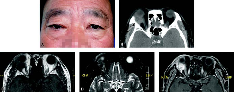
Figure 1 Appearance and imaging of pleomorphic adenocarcinoma of the lacrimal gland
A. External appearance of a patient with pleomorphic adenocarcinoma of the right lacrimal gland, showing right eye proptosis and ptosis of the upper eyelid.
B. Axial CT imaging reveals a hyperdnese extraconal orbital mass with no evidence of bone erosion (black arrow).
C. The tumor appears isointense on T1-weighted MRI.
D. MRI T2-weighted imaging with fat suppression reveals inhomogeneous signals within the tumor.
E. Contrast-enhanced T1-weighted imaging shows inhomogeneous enhancement of the tumor.
Treatment
The treatment principles align with those for adenoid cystic carcinoma of the lacrimal gland.