Pleomorphic adenoma of the lacrimal gland, also known as benign mixed tumor of the lacrimal gland, primarily originates in the orbital portion of the lacrimal gland.
Clinical Presentation
This condition is most commonly observed in individuals between the ages of 40 and 60 and typically involves unilateral symptoms. It progresses slowly and manifests as proptosis with downward displacement of the eyeball. On palpation, a firm, smooth, well-demarcated, and painless mass can often be felt in the superotemporal region of the orbit. CT scans reveal a round or oval, isodense mass in the lacrimal gland region, which may cause bony indentation or enlargement of the lacrimal gland fossa. MRI scans show the tumor with isointense signals on T1-weighted imaging (T1WI), mixed signals on T2-weighted imaging (T2WI), and inhomogeneous enhancement on contrast-enhanced scan. Although pleomorphic adenomas of the lacrimal gland often have a pseudo-capsule, the boundary between the tumor and capsule may not be clearly defined, and nodular protrusions on the surface can be observed. The cut surface of the tumor often reveals not only tumor cell components but also areas of mucinous and fibrous tissue. Under the microscope, the tumor comprises a mixture of glandular epithelial cells, myoepithelial cells, and stromal components.
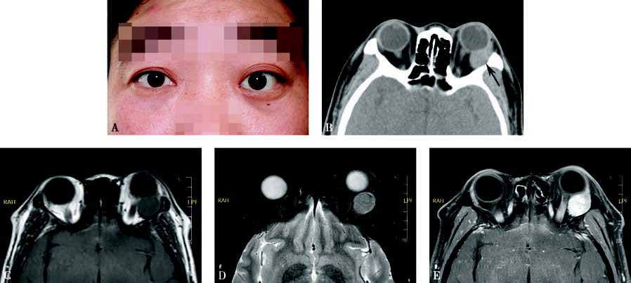
Figure 1 Appearance and imaging of pleomorphic adenoma of the lacrimal gland
A. A photograph of a patient with pleomorphic adenoma of the left lacrimal gland, showing left proptosis and downward displacement of the eyeball.
B. Axial CT imaging shows an isodense mass in the left lacrimal gland region with bony indentation of the lacrimal fossa (black arrow).
C. MRI T1-weighted imaging demonstrates an isointense tumor signal.
D. MRI T2-weighted imaging with fat suppression reveals a tumor with mixed signals.
E. Contrast-enhanced T1-weighted imaging shows inhomogeneous enhancement of the tumor.
Treatment
The primary treatment is surgical resection, with complete removal of the tumor being essential. Residual or ruptured tumor capsule increases the risk of recurrence and potential malignant transformation.