Conjunctival lymphoma can manifest as an isolated lesion or coexist with lymphoma in other organs. There is a significant association between microbial infections and the development of conjunctival lymphoma, and autoimmune diseases are also linked to its occurrence.
The condition often presents unilaterally, with patients typically seeking medical attention due to a sensation of a foreign body in the eye. In the early stages, eyelid swelling and conjunctival edema may be observed. The characteristic clinical feature is an elevated "salmon-pink" appearance of the lesion, which is most commonly located in the conjunctival fornix. As the disease progresses, the lesion may extend along tissue planes to involve the extraconal orbital space, extraocular muscles, and the lacrimal gland, with deeper orbital invasion leading to restricted eye movements or proptosis. Histopathologically, mucosa-associated extranodal marginal zone lymphoma (MALT lymphoma) is the most common subtype.
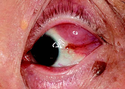
Figure 1 Conjunctival lymphoma in the left eye
The lesion is located in the superotemporal conjunctival fornix, with conjunctival edema presenting as a salmon-pink appearance. Feeder vessels are visible, and the lesion is contiguous with an orbital mass.
Treatment
Treatment involves partial or complete surgical excision of the lesion. Postoperative staging determines the need for additional radiotherapy or chemotherapy.