Conjunctival dermoid tumors and dermolipomas are choristomas and are relatively common congenital benign tumors of the conjunctiva. These are usually sporadic and lack a hereditary pattern. They result from ectopic mature tissue differentiation at the limbus or conjunctiva. The ectopic tissue components may include skin, bone, lacrimal gland, cartilage, and others.
Conjunctival dermoid tumors are most commonly located at the inferotemporal corneal limbus. They appear as round, smooth, yellowish elevated masses, often with fine hair visible on the surface.
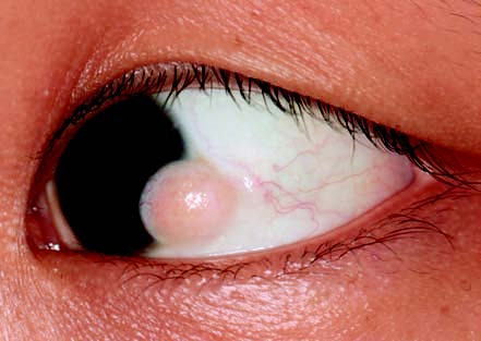
Figure 1 Conjunctival dermoid tumor in the left eye
The lesion is located at the inferotemporal corneal limbus. It is a yellowish-white solid mass with a smooth surface, involving the cornea, and has well-defined borders.
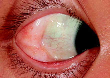
Figure 2 Conjunctival dermolipoma in the right eye
The lesion is located at the lateral canthus of the bulbar conjunctiva. It presents as a yellowish-white, soft mass that is connected to orbital tissue and has relatively well-defined borders.
Conjunctival dermolipomas are typically found beneath the bulbar conjunctiva in the superotemporal quadrant near the lateral canthus. They present as soft, smooth, yellowish masses.
Treatment
Dermoid tumors that significantly involve the cornea may affect visual development, and early surgical intervention may be necessary in such cases. Dermolipomas generally do not require treatment unless the lesion is large or cosmetically concerning. Surgical removal may be considered in these situations, but deep excision should be approached with caution due to the potential risk of visual function-related complications, such as strabismus or ptosis.