Conjunctival papilloma arises from the stratified squamous epithelium of the conjunctiva and is more commonly observed in males, with a peak incidence between the ages of 21 and 40. Human papillomavirus (HPV) infection is the primary risk factor. Subtypes 6 or 11 are associated with the development of pedunculated conjunctival papillomas, while subtypes 16 or 18 are more often linked to broad-based conjunctival papillomas.
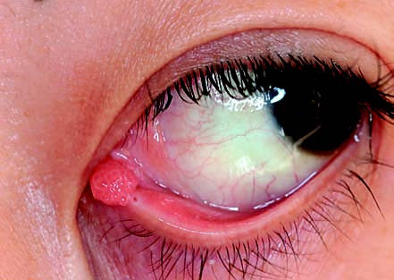
Figure 1 Conjunctival papilloma of the left eye
The lesion is located on the nasal side of the lower punctal conjunctiva. It appears as a pedunculated, flesh-colored papillomatous mass with well-defined borders.
The tumor typically occurs in areas such as the limbus, caruncle, or eyelid margin. It appears as a pale red or flesh-colored, fleshy mass that may be either pedunculated or sessile. Symptoms may include a sensation of a foreign body, dryness, conjunctival bleeding, or abnormal appearance. When the lesion affects the lacrimal duct, it may result in tearing or bloody tears. The surface of the tumor is irregular and often consists of multiple lobules with a smooth appearance and spiral-shaped blood vessels. Pathological examination reveals that the papilloma is composed of multiple lobules, with proliferative squamous epithelium covering vascularized fibrous connective tissue. Acute or chronic inflammatory cell infiltration may also be observed microscopically.
Treatment
The primary treatment is surgical removal.