A conjunctival nevus, derived from the neuroectoderm, is the most common melanocytic tumor of the conjunctiva and is frequently observed in children. It usually occurs near the limbus and on the bulbar conjunctiva within the palpebral fissure area. The lesion appears as an irregular or slightly rounded shape, varies in size, has well-defined borders, and is slightly elevated above the conjunctival surface. The color is typically black or gray-black, although some lesions may appear brownish-red. Vascularization is generally absent within the nevus. A sudden increase in size, the development of a rough surface, or the appearance of vascular infiltration suggests the potential for malignant transformation. Histopathological examination typically shows that conjunctival nevi are composed of nevus cells or nests.
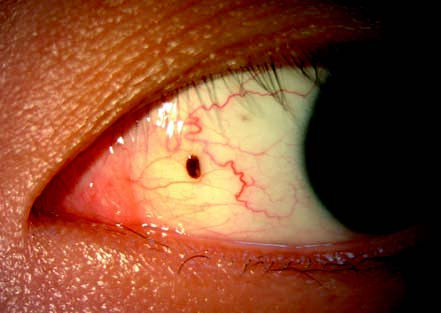
Figure 1 Conjunctival nevus of the left eye
The lesion is located on the nasal side of the bulbar conjunctiva. It is gray-black with clear borders, and histopathology confirms an intradermal nevus.
Treatment
Treatment is generally unnecessary. However, lesions that significantly affect appearance can be removed completely. Pathological examination of the excised tissue is routinely performed, and if malignancy is confirmed, excision with no-touch wide margins is recommended.