Basal cell carcinoma of the eyelid is the most common malignant eyelid tumor. It predominantly affects middle-aged and elderly individuals, with photochemical damage being the most significant causative factor. The tumor commonly arises on the lower eyelid near the inner canthus. Initially, it presents as a small nodule with visible telangiectasia on the surface. The lesion is markedly elevated, firm in texture, and grows slowly. Due to its abundant pigmentation, it may be mistaken for a pigmented nevus or melanoma. Over time, ulceration develops in the central part of the tumor, with undermined, crater-like edges that gradually invade surrounding tissues, causing extensive destruction. Histologically, the tumor consists of small, uniformly shaped, compact lobules of basophilic cells with scant cytoplasm. Basal cell carcinoma rarely metastasizes.
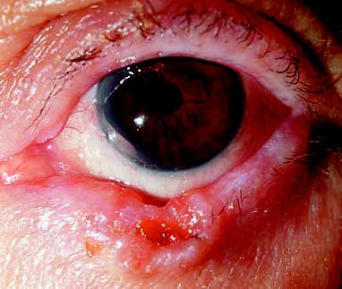
Figure 1 Basal cell carcinoma of the left eyelid margin
The lesion demonstrates raised edges and central ulceration.
Treatment
Surgical excision is the standard treatment. If the lesion is extensive and cannot be completely excised, postoperative radiotherapy or treatment with Hedgehog pathway inhibitors may be utilized.