A pigmented nevus of the eyelid is a congenital flat or elevated lesion composed of nevus cells, with well-defined margins. Pigmentation may be present from childhood or may not appear until puberty or adulthood. Histologically, pigmented nevi can be classified into the following types:
- Junctional Nevus: Nevus cells are located at the dermo-epidermal junction. It appears as a flat pigmented macule, round or oval in shape, with slow growth. There is a low potential for malignant transformation.
- Intradermal Nevus: This is the most common type, often presenting as an elevated lesion, sometimes with a papillomatous appearance. Pigmentation is minimal, if present, ranging from brown to black. Nevus cells are entirely located within the dermis, and it generally has no tendency for malignant transformation.
- Compound Nevus: This is typically brown in color and is a combination of features from the first two types. There is a low risk of malignant transformation.
- Blue Nevus: Often flat, this type is characterized by pigmentation present at birth, appearing blue or slate-gray in color. It has no malignant potential.
- Congenital Eyelid Melanocytosis (also known as Ota Nevus): This is a type of blue nevus involving the periocular skin, including the eyelid. In rare cases, it can undergo malignant transformation, and some patients may develop associated choroidal melanoma.
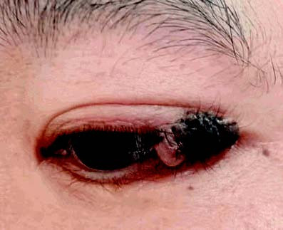
Figure 1 Pigmented nevus at the left eyelid margin
The lesion shows pigmentation and may exhibit hair growth.
Treatment
Pigmented nevi of the eyelid typically require no intervention unless there are indications for removal due to cosmetic concerns or visual obstruction. If signs of malignant transformation are observed, such as rapid growth, deepening pigmentation, ulceration, or bleeding, complete excision with pathological evaluation is necessary.