Eyelid hemangioma is the most common benign tumor of the eyelid during infancy and early childhood. It consists of proliferating capillaries and endothelial cells. The condition typically appears shortly after birth and follows a course generally divided into three phases: the proliferative phase, plateau (stabilization) phase, and involution phase. Rapid growth occurs during the proliferative phase, and the lesion often regresses partially or completely by the age of 6 to 7 years.
Superficial hemangiomas of the eyelid appear bright red in color. Subcutaneous hemangiomas of the eyelid are sometimes connected to orbital hemangiomas, exhibiting a bluish or purplish hue. Such lesions may exert pressure on the eye, leading to astigmatism, anisometropia, strabismus, or amblyopia. Eyelid hemangioma should be distinguished from port-wine stain (PWS), which is a congenital anomaly composed of dilated sinusoidal vessels. Unlike hemangiomas, port-wine stains are flat, progress slowly, and in some cases, are associated with Sturge-Weber syndrome.
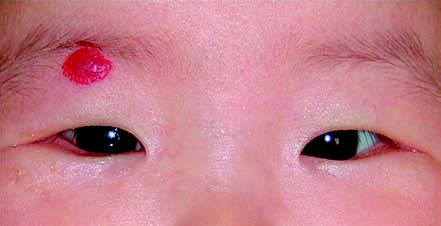
Figure 1 Hemangioma of the right eyelid
The lesion is superficial and presents as a bright red area.
Treatment
Eyelid hemangiomas often show a spontaneous tendency to regress, and smaller lesions may be monitored with regular observation. If the lesion causes ptosis or abnormal positioning of the eye, treatment is warranted. The first-line therapy involves the use of beta-blocker medications.