Orbital fibrous dysplasia is a benign bone lesion caused by somatic mutations in the GNAS gene during embryonic development. This condition is characterized by impaired differentiation and maturation of osteoblasts, where normal lamellar bone is replaced by immature woven bone and fibrous tissue. It can affect any bone in the body, presenting as either monostotic (single bone) or polyostotic (multiple bones) lesions. Typical features include deformities, pain, and pathological fractures.
Clinical Manifestations
Fibrous dysplasia can be classified into the monostotic type, polyostotic type, McCune-Albright syndrome, and Mazabraud syndrome. Polyostotic disease associated with skin pigmentation and endocrinopathies, such as precocious puberty, is referred to as McCune-Albright syndrome. The presence of fibrous dysplasia with intramuscular myxomas defines Mazabraud syndrome.
A painless orbital deformity is the most common reason for seeking medical attention. Visual impairment is one of the frequent ocular manifestations, often caused by compression or traction of the optic nerve by the affected bone. In most cases, visual deterioration is gradual, progressing over months to years, though acute vision loss can occur in rare instances. When the lesion involves the orbit and compresses the globe, symptoms such as proptosis, globe displacement, restricted eye movement, and diplopia may arise. Lesions invading the maxilla can compress the bony nasolacrimal duct, leading to symptoms like epiphora and chronic dacryocystitis.
Extra-skeletal manifestations include jagged-edged café-au-lait macules that do not cross the midline, and abnormalities in hormones such as sex hormones, thyroid hormones, growth hormone, and cortisol. Intramuscular myxomas associated with Mazabraud syndrome commonly present as painless, firm, and palpable masses, typically found in the limbs. The overall malignancy risk for fibrous dysplasia is low (0.4%–4%). However, rapid lesion growth, new onset of bone pain, or a history of radiation therapy should prompt suspicion of potential malignant transformation.
CT imaging is a routine diagnostic tool. It often reveals localized or extensive bone expansion deformities with poorly defined borders. The cortical bone is thinned, and there are expansile changes within the medullary cavity that are irregular in shape with indistinct boundaries. Trabecular bone structure is typically absent, displaying a ground-glass appearance. In children and adolescents, the lesions are usually relatively homogeneous but become less transparent and more heterogeneous with age.
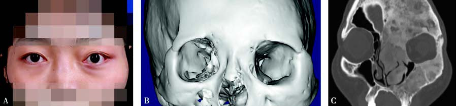
Figure 1 Fibrous dysplasia of the bone
A: Patient’s facial appearance showing midfacial prominence and left-sided proptosis. B: CT 3D reconstruction revealing expansile growth of the left frontal, zygomatic, maxillary, and ethmoidal bones. C: Coronal CT scan demonstrating expansile and ground-glass changes in the left frontal, zygomatic, maxillary, and ethmoidal bones.
Treatment
Asymptomatic patients may not require immediate treatment but necessitate close follow-up, including annual neurological, ophthalmological, and auditory evaluations, as well as regular CT imaging. Pharmacological interventions can temporarily alleviate pain, improve bone density, and reduce the risk of pathological fractures. Surgical treatment remains the most common approach for symptomatic patients or those with functional impairment. Small lesions can be completely resected, while larger and more diffuse lesions are often partially removed to improve appearance and function.
Therapeutic optic nerve decompression is recommended for patients with progressive vision loss caused by optic nerve compression. Prophylactic decompression is not suggested for asymptomatic patients. Due to the destruction of normal structures and challenges in localization, endoscopic navigation technology is recommended to assist in surgical procedures.