Congenital microphthalmia and anophthalmia represent congenital orbital abnormalities resulting from failure of the embryonic fissure to close during embryonic development. This leads to proliferation of neuroepithelial tissue in the orbit, forming a cyst. The inner layer of the cyst consists of dysplastic retina with indistinct structure, and in some cases, only remnants of the scleral shell or traces of ocular adnexa remain.
Clinical Manifestations
Individuals with this condition typically present with a non-functional small eye, sometimes accompanied by a cyst. The cyst is often located below the microphthalmic eye and is connected to it. The lower eyelid tends to appear bulged, with a cystic consistency. The size of the cyst can vary, and it may move along with the eye when ocular movement occurs.
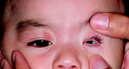
Figure 1 Congenital microphthalmia of the left eye.
Treatment
Management involves the periodic replacement of ocular prostheses to stimulate orbital development and reduce the risk of facial asymmetry.