Thyroid eye disease (TED) is a chronic, multisystem disorder caused by an autoimmune reaction and is closely associated with thyroid dysfunction. It is the most common orbital disease in adults and is also known as Graves’ ophthalmopathy (GO) or thyroid-associated ophthalmopathy (TAO). Epidemiological studies from the United States estimate an annual incidence of about 16 per 100,000 women and 2.9 per 100,000 men.
Etiology
The pathogenesis of TED is not fully understood. It is primarily related to autoimmunity, with contributions from genetic and environmental factors. When the immune system fails to maintain tolerance to thyrotropin receptor (TSHR), B cells and plasma cells produce autoantibodies that target orbital fibroblasts. Concurrently, helper T cells (Th1, Th2, and Th17) release cytokines such as interferon-γ, interleukin-4, and interleukin-17a, respectively, which synergistically stimulate the proliferation and differentiation of orbital fibroblasts into adipocytes and myofibroblasts. These processes lead to the characteristic pathological changes in TED: tissue edema, adipose tissue hyperplasia, and fibrosis.
Clinical Manifestations
TED affects a wide range of orbital structures, including the eyelids, lacrimal glands, extraocular muscles, orbital fat, connective tissues, and the ocular surface. Secondary complications contribute to its highly varied and complex clinical presentation. Common symptoms include photophobia, tearing, dryness, a sensation of a foreign body, proptosis, restricted ocular motility, and diplopia. Severe proptosis combined with eyelid retraction can lead to incomplete eyelid closure, resulting in exposure keratitis, corneal ulcers, or even perforation. Significant enlargement of the extraocular muscles may compress the optic nerve, causing optic neuropathy, impaired color vision, reduced visual acuity, and potentially blindness. The bilateral involvement of both eyes is more common, although the presentation may differ between the two eyes and may occur sequentially.
Clinically, TED is categorized into two primary forms. In the first group, hyperthyroidism coincides with ocular symptoms, presenting prominently with orbital inflammation, which is primarily characterized by adipose tissue edema on imaging. Extraocular muscle enlargement is less pronounced, and orbital soft tissue fibrosis occurs relatively late. This form is more common in adult females, responds well to corticosteroids, but tends to recur. In the second group, mild or normal thyroid function is observed at the time of ocular disease onset. In this form, extraocular muscle enlargement is the predominant feature on imaging, while orbital adipose tissue edema and hyperplasia are minimal. Early fibrosis of orbital soft tissues may occur. This type is more common in adult males and is less responsive to corticosteroid therapy.
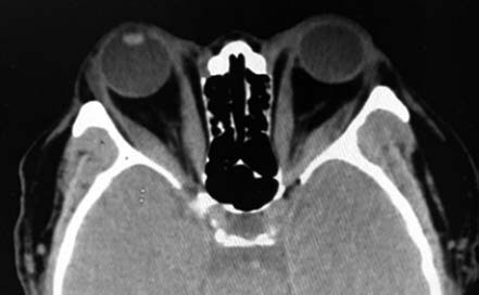
Figure 1 Thyroid eye disease
CT imaging shows bilateral proptosis with fusiform thickening of the medial rectus muscles.
Based on disease progression, TED is divided into an active phase and an inactive phase. Assessing disease activity is critical for determining the timing of treatment and estimating prognosis. The clinical activity score (CAS) is commonly used for evaluating inflammation in the eyelids, conjunctiva, and caruncle, and imaging studies such as MRI may be employed for further assessment. Depending on the severity of clinical symptoms, TED can be classified as mild, moderate-to-severe, or very severe. Patients with exposure keratitis or optic neuropathy are categorized as having very severe TED.
Major Ocular Manifestations
Eyelid Changes
Eyelid retraction and lag are key features of TED due to involvement of the levator palpebrae superioris muscle and Müller’s muscle. Eyelid retraction is characterized by exposure of the sclera above the limbus when the eyes are open, known as Dalrymple’s sign. Eyelid lag occurs when the upper eyelid fails to follow the downward movement of the globe, exposing the upper sclera, referred to as von Graefe’s sign.
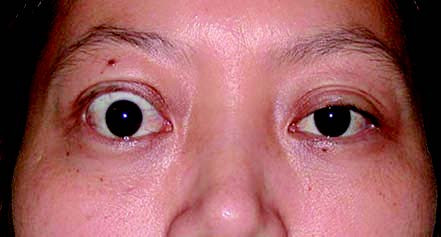
Figure 2 Thyroid eye disease
Right upper eyelid retraction demonstrating Dalrymple’s sign.
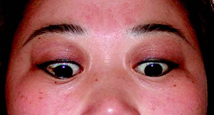
Figure 3 Thyroid eye disease
Bilateral upper eyelid lag on downgaze, corresponding to von Graefe’s sign.
Eyelid and Conjunctival Inflammation
Involvement of the eyelids leads to congestion and edema. Increased orbital pressure from intraorbital tissue edema causes conjunctival congestion and edema, which can be severe enough to result in conjunctival prolapse through the palpebral fissure.
Proptosis
Proptosis is typically bilateral but may occur sequentially. In early stages, axial proptosis is common. In later stages, fibrosis and contraction of the extraocular muscles may cause proptosis along with fixation in a specific eye position. A subset of patients develops worsening proptosis after treatment of hyperthyroidism, referred to as malignant proptosis.
Ocular Motility Disorders and Diplopia
Extraocular muscle involvement is common, leading to restrictions in ocular motility and diplopia. The involvement frequency of muscles follows the order: inferior rectus, medial rectus, superior rectus, and lateral rectus. CT imaging typically shows thickening of the muscle belly with sparing of the tendinous insertions, which helps distinguish TED from idiopathic orbital myositis. Fibrosis of the extraocular muscles exacerbates motility issues and diplopia. For patients with concurrent eyelid closure insufficiency, restricted motility further increases the risk of corneal damage.
Corneal and Ocular Surface Disease
Severe proptosis and eyelid retraction can lead to incomplete eyelid closure. Autoimmune inflammation involving the ocular surface can result in keratoconjunctivitis, corneal ulcers, or even corneal perforation in advanced cases. Symptoms include significant pain, photophobia, and tearing.
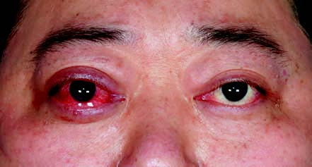
Figure 4 Thyroid eye disease
Bilateral proptosis with right corneal ulceration.
Optic Neuropathy
Intraorbital edema, elevated orbital pressure, and enlargement of extraocular muscles can compress the optic nerve, causing optic neuropathy. Manifestations include afferent pupillary defects, color vision deficiencies, visual field defects, and reduced visual acuity. In severe cases, only light perception may remain. Fundus examination may reveal optic disc edema or pallor, retinal edema, and venous engorgement.
Intraocular Pressure Elevation
Orbital edema, increased retrobulbar fat, and thickened extraocular muscles contribute to crowded orbital spaces, elevated orbital pressure, and increased intraocular pressure, typically less than 30 mmHg. Systemic symptoms of hyperthyroidism, such as thyroid enlargement, irritability, increased basal metabolic rate, tachycardia, weight loss, increased appetite, hand tremors, and pretibial myxedema, are often present in affected patients.
Diagnosis
The diagnosis of TED is generally not difficult and is primarily based on the following three aspects:
- Typical ocular signs and symptoms, such as eyelid retraction, proptosis, strabismus, and diplopia.
- Abnormal thyroid function or thyroid-related antibodies.
- Imaging findings, including fusiform thickening of extraocular muscles.
Treatment
The treatment of TED includes pharmacological therapy, orbital radiation therapy, and surgical intervention. It also requires ongoing management of risk factors, stabilization of thyroid function, and supportive care for ocular symptoms.
Corticosteroid therapy is one of the main treatment modalities for active-phase TED. It helps alleviate inflammatory responses, shortens the duration of the active phase, and facilitates the transition to the inactive phase. Corticosteroids can be administered intravenously, orally, or locally. Intravenous high-dose corticosteroid pulse therapy is the first-line treatment for moderate-to-severe active-phase TED. A total dose of 4.5 g methylprednisolone is recommended, with 500 mg administered weekly via intravenous infusion for six weeks, followed by 250 mg weekly for another six weeks.
Biological agents are emerging treatment options. Targeted therapies such as teprotumumab, rituximab, and tocilizumab can be considered as second-line treatments.
For patients who are contraindicated for corticosteroid therapy or are unresponsive to hormones, orbital radiation therapy may be employed. This approach is suitable for patients with muscular hypertrophy in the active phase of TED, typically involving bilateral temporal field irradiation with a total dose of approximately 20 Gy.
In the inactive phase of TED, surgical correction may be performed if proptosis, strabismus, or eyelid deformities impair vision, quality of life, or aesthetic appearance. Emergency surgery is also indicated for severe exposure keratitis or optic nerve compression that cannot be adequately treated with non-surgical measures. The primary surgical procedures include orbital decompression, extraocular muscle surgery, and eyelid surgery, which are often conducted in stages:
- Orbital decompression surgery to reduce proptosis, relieve exposure keratitis, and alleviate optic nerve compression.
- Extraocular muscle surgery to correct strabismus and improve diplopia.
- Eyelid surgery to address eyelid retraction and improve cosmetic appearance.
Recent advancements in orbital surgical navigation, endoscopy, and robotic technologies have significantly enhanced the safety and effectiveness of these procedures.