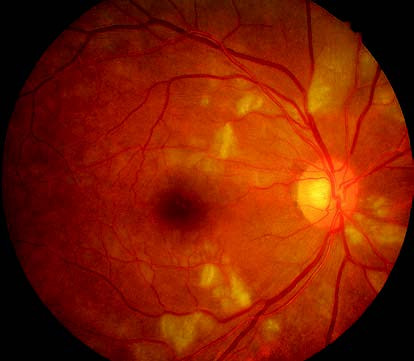
Precapillary retinal arteriolar occlusion manifests as irregularly shaped, variably sized, and poorly defined grayish-white patchy lesions on the superficial retina, commonly referred to as cotton-wool spots. These lesions represent localized ischemic infarctions of the retinal nerve fiber layer. They are generally smaller than one-quarter of the optic disc area and typically resolve within 5 to 7 weeks, although the duration may be longer in diabetic patients. This condition is commonly observed in individuals with diabetic retinopathy (DR), hypertensive retinopathy, nephropathy-associated retinopathy, systemic lupus erythematosus, leukemia, or acquired immune deficiency syndrome (AIDS), among other systemic diseases. When cotton-wool spots are detected in the fundus, systemic causes should be investigated. Approximately 95% of cases are associated with an underlying serious systemic disease.

Figure 1 Precapillary retinal arteriolar occlusion in the right eye (Cotton-Wool spots)
Yellowish-white, spot-like lesions are scattered on the superficial retina.