Etiology
The causes are similar to those of CRAO, with embolism and inflammation being the primary reasons. Embolic sources are the same as in CRAO and include cardiogenic emboli, carotid or aortic-origin emboli, and fat emboli. The most common type is the cholesterol embolus, which often originates from atherosclerotic plaques in the carotid artery. Calcified emboli are typically larger than cholesterol emboli, often originating from heart valves, and are more likely to cause more severe obstructions.
Clinical Manifestations
Visual acuity may show varying degrees of reduction, and a fixed scotoma can be present in a specific area of the visual field. On fundoscopy, the affected branch artery appears narrowed, and the retina in the area supplied by the occluded artery shows grayish-white edema. The retinal edema is most prominent in the posterior pole along the course of the obstructed vessel. Emboli can sometimes be seen within the occluded branch artery.
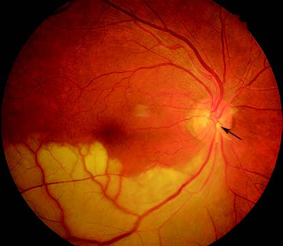
Figure 1 Color fundus photography of branch retinal artery occlusion (BRAO) in the right eye
Occlusion of the inferotemporal retinal artery in the right eye is evident, with a visible embolus (black arrow) in the artery. The retina in the territory supplied by the occluded artery shows grayish-white edema.
Treatment
Systemic causes should be identified and treated accordingly. Other management approaches are the same as those for CRAO.