After-cataract refers to the opacification that arises from residual cortical material or proliferated lens epithelial cells following extracapsular cataract extraction (including phacoemulsification) or lens trauma. Posterior capsular opacification, which occurs beneath the posterior lens capsule after cataract surgery, is the most common complication of extracapsular cataract extraction.
Etiology
Lens epithelial cells that persist beneath the capsule following extracapsular cataract extraction can proliferate and form Elschnig’s pearl-like bodies. These epithelial cells may undergo myofibroblastic differentiation, which leads to contraction and fine folding of the posterior lens capsule. Residual cortical material from incomplete cortical absorption after cataract extraction or traumatic cataract can exacerbate opacification, resulting in visual distortion and reduced visual acuity.
Clinical Features
The incidence of posterior capsular opacification can reach 30%–50% within three or more years after extracapsular cataract extraction. Nearly all pediatric cataract surgeries result in posterior capsular opacification. The posterior lens capsule may show unevenly thickened fibrotic tissue and Elschnig’s pearls. The degree of vision impairment is related to the extent and thickness of the posterior capsular opacification.
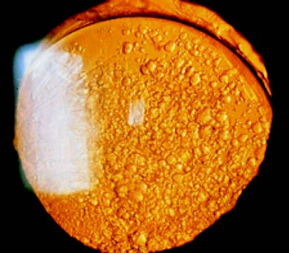
Figure 1 Formation of Elschnig's pearl-like bodies and opacification of the posterior capsule after cataract extraction
Diagnosis
A history of extracapsular cataract extraction or lens trauma provides context. The use of a slit-lamp microscope enables an accurate assessment of the presence and degree of posterior capsular opacification.
Treatment
When after-cataract affects visual acuity, a Nd:YAG laser can be used to create an opening in the posterior capsule within the pupillary zone. In cases where laser treatment is unavailable or the opacification is too dense for laser application, surgical incision or excision of the posterior capsule in the pupillary zone may be performed. Postoperative management may involve the use of topical glucocorticoids or nonsteroidal anti-inflammatory eye drops to prevent inflammatory reactions, alongside monitoring of intraocular pressure for potential changes.