Meibomian gland dysfunction (MGD) is a chronic, nonspecific inflammatory condition of the meibomian glands, characterized by obstruction of the gland ducts or abnormal glandular secretion. It is the primary cause of evaporative dry eye.
Etiology
The pathogenesis of MGD remains incompletely understood but is thought to be associated with degenerative changes in the meibomian glands. Its occurrence is closely related to certain dermatological conditions, such as rosacea, seborrheic dermatitis, atopic dermatitis, psoriasis, and systemic lupus erythematosus. Studies have indicated a strong association between Demodex mite infestation and MGD. The compositional abnormality of meibum secreted by the meibomian glands, including elevated cholesterol esters and free fatty acid esters, promotes the growth of Staphylococcus aureus and leads to blepharitis. Lipases and esterases produced by coagulase-negative staphylococci, Propionibacterium acnes, and Staphylococcus aureus can degrade the meibum secreted by the glands, producing fatty acids and triglycerides that are released into the tear film. This creates foam, destabilizes the tear film, and can irritate the eyelids, exacerbating ocular discomfort. In advanced cases, gland atrophy, loss of acini, keratinization, and scarring of the gland ducts may occur.
Clinical Manifestations and Examinations
MGD is common in older adults and is more frequent in individuals with oily skin. It often co-occurs with blepharitis. There is no significant gender difference in prevalence, but colder regions exhibit a higher incidence compared to warmer climates. The main symptoms include a burning sensation in the eyes, foreign body sensation, dryness, irritation, and visual fatigue. Common clinical signs include thickened eyelid margins, which may present with erythema and hyperkeratosis, as well as vascular dilation on the posterior eyelid margin extending from posterior to anterior. Meibomian gland orifices may be blocked by white keratotic material, which can appear raised and deformed. Expression of the glands may yield foamy, granular, or toothpaste-like secretions. With disease progression, yellowish mucoid secretions may develop, and chronic inflammation over years can lead to widespread atrophy of the meibomian glands. Other associated signs often observed include meibomian cysts, conjunctival concretions, conjunctival hyperemia, papillary hypertrophy, and punctate corneal staining. Severe cases may lead to corneal pannus, corneal ulcers, or ectropion. Examinations for dry eye may reveal tear film deficiency, instability, and increased tear osmolarity.
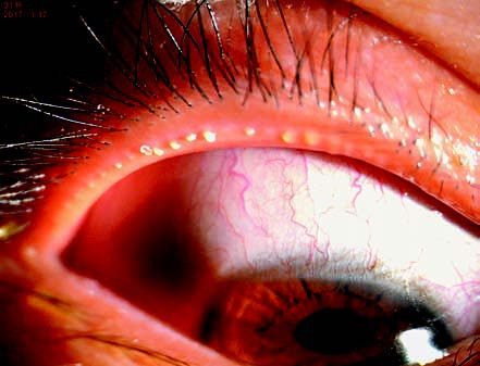
Figure 1 Meibomian gland dysfunction
The meibomian gland orifices exhibit blockage by white keratinized material, appearing raised and deformed. Upon expression, the secretions present a toothpaste-like consistency.
The eyelid margin and meibomian glands are critical components of the lacrimal functional unit. Assessing alterations in the eyelid, lid margin, and meibomian glands provides valuable insight into the classification of dry eye.
Abnormalities of the Lid Margin
Patients with MGD may show thickening, blunting, or irregularity of the lid margin, along with hyperemia or the development of neovascularization. "Collarette signs" (cylindrical dandruff around the base of eyelashes) are suggestive of Demodex infestation, and further mite-related evaluation may be warranted.
Evaluation of Meibomian Gland Morphology and Function
Morphological and functional assessment of the meibomian glands is a routine step in diagnosing MGD. In addition to meibography for structural assessment, evaluation can include observation of the gland orifices and assessment of the ease with which meibum is expressed upon manual compression.
Diagnosis
Diagnosis of MGD primarily focuses on two key clinical features: meibomian gland orifice condition and meibomian secretion status.
(1) Abnormalities in gland orifices: These may include obstruction, narrowing, displacement, closure, or congenital absence of the orifices.
(2) Meibomian secretion abnormalities: These involve impaired glandular expression and/or altered characteristics of secreted meibum.
The presence of either (1) or (2) is sufficient for a diagnosis of MGD.
For patients exhibiting any of the above abnormalities without accompanying clinical symptoms, the condition is diagnosed as asymptomatic MGD. Early identification and initiation of treatment in such cases can help slow disease progression and prevent further deterioration.
Treatment
Treatment Principles
Treatment is aimed at eliminating or reducing causative factors, preventing risk factors, restoring the function of the meibomian glands, improving the ocular surface microenvironment, inhibiting inflammatory responses, and preventing complications.
Treatment Approaches
Physical Therapy
Application of warm compresses, eyelid hygiene, and meibomian gland massage can be seen in the management approaches for dry eye.
Intense pulsed light (IPL) primarily alleviates eyelid margin inflammation and relieves symptoms and signs of MGD and related dry eye disease through its thermal and antimicrobial effects. Thermal pulsation therapy involves concurrent heating of the eyelids and pulsatile massage of the meibomian glands.
Pharmacological Therapy
Artificial tears can be seen in the management approaches for dry eye.
Topical antimicrobials and anti-Demodex agents should be used when bacterial infection or Demodex infestation is clearly identified. Eye gels or ointments are commonly applied to the eyelid margins. Frequently used antimicrobial agents include tetracyclines, macrolides, quinolones, and aminoglycosides. Common anti-Demodex agents include metronidazole, tea tree oil, and okra-based medications.
Topical anti-inflammatory therapy can be seen in the management approaches for dry eye.
In severe cases of MGD, systemic antibiotics can be administered, utilizing their anti-inflammatory properties rather than their antibacterial effects. These include tetracyclines and macrolides. For patients with systemic inflammatory dermatoses such as rosacea, seborrheic dermatitis, or acne rosacea, collaboration with dermatology is recommended.
Other Therapies
Improvement of Dietary Habits
Reducing intake of high-sugar, high-fat, spicy, and irritant foods is beneficial. Increased consumption of omega-3 fatty acids has been shown to improve tear secretion function and alleviate MGD symptoms.
Adjunctive Treatments and Functional Training
Incomplete blinking leads to impaired glandular function and can contribute to MGD. Training to promote complete blinking is recommended for such individuals. The approach includes encouraging patients to completely close their eyes for 2–3 seconds before reopening them, with daily practice of 100–200 repetitions.
Surgical Interventions
Meibomian gland duct probing combined with physical or pharmacological treatments has demonstrated efficacy in improving MGD symptoms and signs and is suitable for patients who do not respond to conventional therapies.