Granular corneal stromal dystrophy is a type of corneal stromal dystrophy that follows an autosomal dominant inheritance pattern. It is caused by a mutation (p.Arg555Trp) in the transforming growth factor beta-induced (TGFBI) gene located on chromosome 5q31.
Pathologically, it is characterized by distinctive features. The corneal granules consist of hyaline material, although the exact nature and origin of these granules remain unclear. They are thought to possibly result from abnormalities in the synthesis or metabolism of cell membrane proteins or phospholipids.
Clinical Manifestations
Onset typically occurs between the ages of 10 and 20, but symptoms may remain absent for many years. The condition develops symmetrically in both eyes and becomes more pronounced after puberty. In addition to varying degrees of visual impairment, other symptoms may not be present. When corneal epithelial erosions occur, redness and photophobia may develop. Grayish-white punctate opacities can be observed beneath the anterior limiting lamina in the central cornea. These opacities vary in size, are sharply demarcated, and appear as round or irregularly shaped deposits with diverse morphologies. They gradually extend into the deeper layers of the corneal stroma, while the intervening corneal tissue remains completely normal.
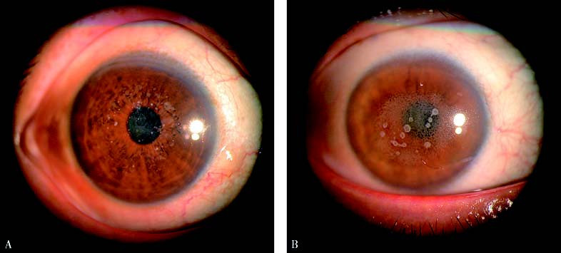
Figure 1 Granular corneal stromal dystrophy
A. Grayish-white punctate opacities of varying sizes with well-defined borders are visible beneath the anterior limiting lamina in the central cornea, with the intervening cornea appearing completely normal.
B. Grayish-white opacities of varying sizes with well-defined borders, presenting as round or irregularly shaped deposits, are visible beneath the anterior limiting lamina in the central cornea, with the intervening cornea appearing completely normal.
Treatment
In the early and intermediate stages, treatment may not be necessary. When significant vision loss affects work and daily life, corneal transplantation or phototherapeutic keratectomy (PTK) with an excimer laser may be considered. However, there is a potential for recurrence after surgery.