Epithelial basement membrane dystrophy (EBMD), also known as map-dot-fingerprint dystrophy, is the most common type of anterior corneal dystrophy. Histopathological examination shows abnormal proliferation of the basement membrane, with projections of the abnormal basement membrane into the epithelium. Epithelial cells may lack hemidesmosomes, and cysts are often present within the epithelium, typically located beneath the basement membrane. These cysts contain cellular debris and nuclear fragments. Most cases do not follow a hereditary pattern and are simply considered degenerative changes. A small proportion may exhibit autosomal dominant inheritance and present as familial cases.
Clinical Manifestations
The disease has an estimated prevalence of about 5%, with a higher incidence in females, and typically affects both eyes. The main symptoms include recurrent spontaneous eye pain, irritation, and temporary visual blurriness. Grayish-white spots or patches, map-like patterns, and fine fingerprint-like lines can be observed in the central corneal epithelium and basement membrane. Recurrent epithelial erosions may occur.
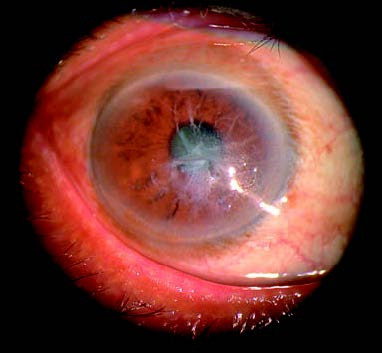
Figure 1 Map-dot-fingerprint dystrophy
Grayish-white spots or patches, map-like patterns, and fine fingerprint-like lines can be observed in the central corneal epithelium and basement membrane, with epithelial erosion present.
Treatment
Treatment involves the use of artificial tears or other viscous lubricants. In cases of epithelial erosion, soft contact lenses may be used, or the epithelium may be debrided, followed by pressure bandaging. For some patients, excimer laser removal of eroded epithelial tissue can promote healing of the new epithelium with satisfactory outcomes.