Band-shaped keratopathy refers to superficial corneal calcific degeneration, primarily affecting the anterior elastic lamina. It is often secondary to various ocular or systemic diseases. It is commonly associated with chronic uveitis, hypercalcemia caused by conditions such as hyperparathyroidism, elevated serum phosphorus with normal calcium levels (as seen in chronic renal failure), and prolonged exposure to mercury-containing agents or solutions (such as long-term use of certain mercury-containing eye drops).
Clinical Manifestations
In the early stages, symptoms are absent. The condition begins with fine, grayish-white calcium deposits in the anterior elastic lamina near the peripheral cornea of the interpalpebral zone. A clear corneal separation is present between the outer edge of the lesion and the limbus, while the inner edge develops a flame-like pattern that gradually extends toward the central cornea. The deposits merge into a band-shaped opacity crossing the interpalpebral zone. When this band crosses the pupillary area, vision declines. The calcium deposits eventually form white, plaque-like lesions that often protrude above the corneal epithelial surface, potentially leading to epithelial defects, irritation symptoms, and a foreign body sensation. In some cases, neovascularization may be observed.
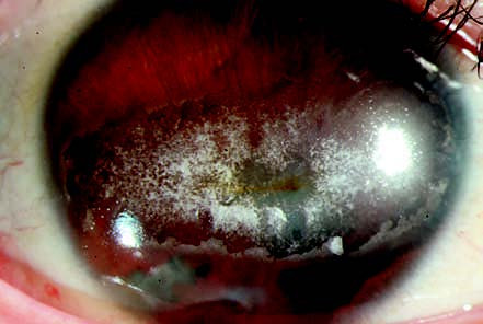
Figure 1 Band-shaped keratopathy
A grayish-white band-shaped opacity is visible across the central cornea, spanning the interpalpebral zone.
Treatment
Treatment of the underlying disease is important. For mild cases, topical disodium edetate (EDTA) eye drops are used. In severe cases, surface anesthesia is applied, followed by epithelial debridement and irrigation of the cornea with 2.5% disodium edetate solution to remove the calcium via chelation. The use of contact lenses soaked in disodium edetate solution may also yield favorable results. In cases with significant opacification, treatment options include lamellar keratoplasty or phototherapeutic keratectomy (PTK) with excimer laser.