Superficial punctate keratitis (SPK) refers to an epithelial lesion of the cornea with an unknown etiology. The condition is unrelated to infection and represents an active inflammation of the cornea without inducing corneal neovascularization.
Clinical Manifestations
This condition can occur at any age, although it is more commonly observed in middle-aged and young adults. Some patients may experience a foreign body sensation, photophobia, and mild vision decline, accompanied by or without mild conjunctival hyperemia. The corneal epithelium demonstrates scattered, small, round or oval nodular or grayish punctate opacities, usually located in the central cornea or visual axis. These lesions are centrally elevated, protruding above the epithelial surface, and show positive staining with fluorescein and rose bengal. The condition may be accompanied by epithelial and subepithelial edema, although no infiltrates are observed.
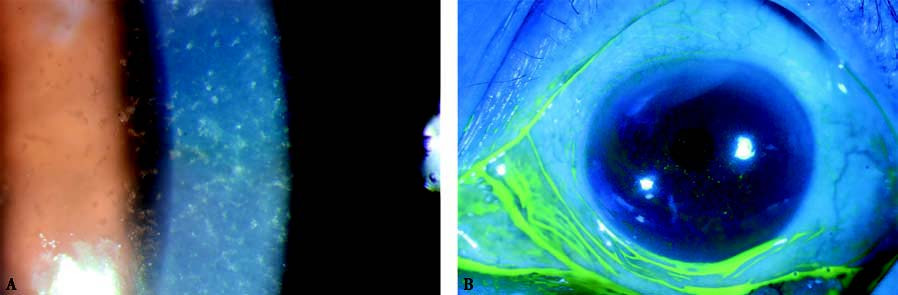
Figure 1 Superficial punctate keratitis
A. The central corneal epithelium shows small, round, nodular or grayish punctate opacities.
B. Fluorescein staining is positive, with lesions appearing as punctate elevations protruding above the corneal epithelial surface.
The corneal epithelium near the lesion may display radial or dendritic appearances, which can occasionally lead to misdiagnosis as herpes simplex keratitis. The lesions can resolve spontaneously within 1 to 2 months without treatment, but they tend to recur after a certain period (typically 6 to 8 weeks). During the remission phase, corneal epithelial defects completely disappear, but mild residual epithelial haze may occasionally persist.
Treatment
In cases of severe acute symptoms, the application of low-concentration corticosteroids shows good therapeutic effects. However, corticosteroids should be used in low doses and for short durations. Therapeutic contact lenses may also be utilized. Medications such as autologous serum, fibronectin, sodium hyaluronate, and growth factors can aid in protecting and promoting the repair of the corneal epithelium. Vitamin supplementation may further support the healing process.
Thygeson’s Superficial Punctate Keratitis
Thygeson’s superficial punctate keratitis is a form of superficial punctate keratitis with an unclear etiology, potentially linked to viral infections and considered an immune response of the host to latent viral infection.
Thygeson’s superficial punctate keratitis mainly manifests as round or oval epithelial opacities with granular white or gray-white appearances. The lesions measure 0.1–0.5 mm in diameter, are slightly elevated, and number between 1 and 20. Minimal or no fluorescein staining is typically observed. These epithelial opacities can occur at any location on the cornea but are most commonly present in the pupillary zone.
The condition fluctuates in severity, alternating between exacerbations and remissions, and may persist for several months or even years. During remission, the lesions that appear in the exacerbation phase may reduce or disappear, only to recur with subsequent exacerbations. Over time, the lesions tend to resolve completely without leaving scars. Corneal sensation generally remains normal, and there is no associated conjunctival hyperemia or corneal edema. Treatment is similar to that for superficial punctate keratitis.