Filamentary keratitis refers to the presence of filamentous structures on the corneal surface, which are composed of degenerated epithelial cells and mucus.
This condition can result from various causes. It is characterized by severe clinical symptoms, challenging treatment, and a tendency for recurrence.
Clinical Manifestations
Symptoms include a foreign body sensation, photophobia, and tearing. The symptoms worsen with blinking but may improve when the eyes are closed. Filamentous structures can be observed on the cornea, with one end adhered to the corneal epithelial surface and the other end free. These filaments can be displaced, and their lengths vary from 0.5 mm to several millimeters. Subepithelial grayish-white opacities may appear beneath the attachment sites of the filaments. Filaments are usually firmly adhered to the cornea, and blinking may cause them to bend or fold. Forceful eyelid closure may result in the detachment of filaments from the corneal surface, leaving behind epithelial defect areas. New filaments may subsequently form in these defect areas, and filaments can appear repeatedly in different locations.
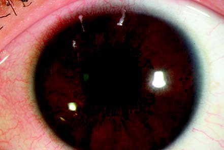
Figure 1 Filamentary keratitis
Curled filamentous structures can be observed in the upper-central region of the cornea, with one end attached to the corneal epithelium and the other end free.
Treatment
Treatment is directed towards addressing the underlying cause. When the sensation of a foreign body caused by the filaments is severe, the filaments can be mechanically removed under topical anesthesia, followed by the use of a bandage contact lens to alleviate symptoms. Antibiotic eye drops are applied to prevent infection. Non-preservative artificial tears and medications that protect the corneal epithelium can be used topically.