Interstitial keratitis is a non-suppurative inflammation of the corneal stroma characterized by cellular infiltration and vascularization, typically without involvement of the corneal epithelium or endothelium. A delayed-type hypersensitivity reaction to infectious agents is associated with the pathogenesis of this condition. Congenital syphilis is the most common cause, while tuberculosis, herpes simplex, herpes zoster, leprosy, mumps, and other infections can also lead to this disease.
Clinical Manifestations
Syphilitic interstitial keratitis is the most common late manifestation of congenital syphilis, predominantly occurring during adolescence (ages 5–20 years).
The disease typically begins unilaterally and often progresses to involve both eyes within weeks to months. It is more frequently observed in females than in males. Early symptoms include ocular discomfort, tearing, photophobia, and significant vision loss.
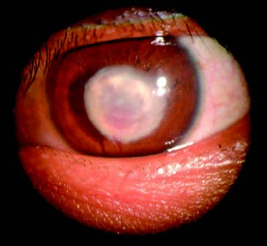
Figure 1 Interstitial keratitis
The central region of the cornea exhibits a near-circular opacity, with stromal neovascularization observed in the deep layers. The blood vessels appear as reddish, brush-like structures between the corneal lamellae.
During the early stages, classical fan-shaped or diffuse corneal stromal infiltration is observed, which may or may not be accompanied by keratic precipitates (KPs). With disease progression, stromal neovascularization develops in deep layers of the cornea, presenting as reddish brush-like vessels between the lamellar layers. The inflammation eventually extends to the central cornea, leading to corneal opacification and edema.
Following resolution of inflammation, edema subsides, but some patients may be left with variably thick corneal scars. Atrophic blood vessels within the stroma appear as fine grayish-white filaments known as ghost vessels. In addition to interstitial keratitis, congenital syphilis commonly presents with other signs such as Hutchinson's teeth, saddle nose, mouth corner cracks, and saber shins. Serological tests for syphilis and specific Treponema pallidum antibody assays aid in diagnosis.
Interstitial keratitis caused by acquired syphilis is less common, generally affecting only one eye, with milder inflammatory responses compared to congenital cases. Typically, it involves a localized quadrant of the cornea and may be accompanied by anterior uveitis.
Tuberculous interstitial keratitis is rare and usually unilateral. It affects specific parts of the cornea, presenting with grayish-yellow plaque-like or nodular stromal infiltrates in the middle to deep layers of the stroma, accompanied by branching neovascularization. The disease is often chronic, with recurrent episodes, and thick scars may remain in the cornea during late stages.
Other types of interstitial keratitis occur in association with conditions such as Cogan's syndrome (characterized by vertigo, tinnitus, hearing loss, and interstitial keratitis), varicella-zoster virus infection, Epstein-Barr virus infection, mumps, rubella, Lyme disease, lymphogranuloma venereum, and onchocerciasis.
Treatment
Systemic therapy involves treatment targeting syphilis or tuberculosis. During the acute inflammatory phase, local administration of cycloplegic agents and corticosteroids is employed to reduce inflammation and prevent complications such as posterior synechiae of the iris and secondary glaucoma. Patients experiencing severe photophobia are advised to use dark-tinted glasses to minimize light exposure. For those with visual impairment caused by corneal scarring, corneal transplantation can be considered.