Pinguecula refers to a horizontal, triangular, or oval-shaped, raised grayish-yellow nodular lesion on the bulbar conjunctiva at the limbal region in the palpebral fissure area.
Etiology
Pathological examination demonstrates hyaline degeneration of the connective tissue beneath the epithelium of the pinguecula, along with an increase in basophilic elastic fibers and granular material. Usually, no inflammatory cells are present in the affected area. This condition is thought to result from ultraviolet (UV) radiation-induced degeneration of collagen.
Clinical Manifestations
The lesion more frequently occurs nasally and tends to develop earlier on the nasal side than on the temporal side. It is typically bilateral. Its appearance is often likened to the infiltration of lipid-like material into the subepithelial tissue and is composed of yellowish translucent elastic tissue. The triangular elevation of the lesion occurs on the bulbar conjunctiva near the limbus, with the triangular base facing the cornea. Pinguecula is usually asymptomatic, with its primary impact being cosmetic. Occasionally, the lesion may become hyperemic, its surface roughened, leading to pingueculitis.
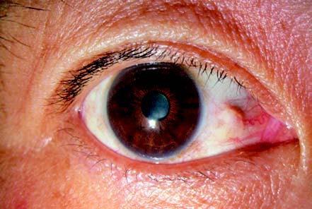
Figure 1 Pinguecula
A yellowish-white nodular elevation is shown on the nasal side, adjacent to the corneal limbus.
Treatment
Treatment is generally unnecessary. If pingueculitis occurs, topical weak corticosteroids or nonsteroidal anti-inflammatory drugs can be administered to relieve inflammation. Lesions that significantly affect cosmetic appearance, are associated with recurrent chronic inflammation, or interfere with successful contact lens fitting, may be considered for surgical excision.