The orbital portion of the lacrimal gland is situated within the lacrimal fossa, suspended by ligaments formed from fascial connective tissue anchored to the orbital periosteum. When these suspensory ligaments become lax, the lacrimal gland dislocates from the lacrimal fossa and descends into the subcutaneous tissue of the temporal upper eyelid, resulting in dislocation of the lacrimal gland.
Etiology
Dislocation of the lacrimal gland is primarily caused by laxity of the suspensory ligaments of the lacrimal gland and often occurs bilaterally. Patients with dermatochalasis frequently exhibit this condition.
Clinical Manifestations
A painless, soft, and easily movable mass may be palpable beneath the skin of the superolateral eyelid. During downward and inward gaze or upon eversion of the upper eyelid, a bulging mass with a pale pink color is visible beneath the superotemporal conjunctiva.
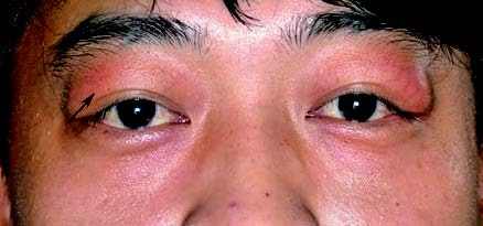
Figure 1 Bilateral dislocation of the lacrimal gland
Prominent masses are observable in the superolateral corners of both eyelids.
Treatment
The condition is managed with lacrimal gland repositioning surgery. This procedure involves suturing the dislocated lacrimal gland back to the periosteum of the lacrimal fossa, combined with reinforcement of the orbital septum.