Acute dacryoadenitis is clinically uncommon and typically occurs unilaterally. It is more frequently observed in children and young adults and often develops as a complication of measles, epidemic mumps, or influenza.
Etiology
The condition is most commonly caused by bacterial or viral infections. The predominant pathogens include Staphylococcus aureus and Neisseria gonorrhoeae. The infection may spread directly from purulent inflammation in the eyelid, conjunctiva, orbit, or face. Alternatively, it can arise from distant purulent foci via hematogenous spread or result from systemic infections.
Clinical Manifestations
Acute dacryoadenitis presents with redness, swelling, and pain in the outer aspect of the upper eyelid, accompanied by tearing. The edematous upper eyelid droops, forming a transverse "S"-shaped curve. Lifting the upper eyelid reveals conjunctival congestion and swelling in the superotemporal region, with mucous discharge. The lacrimal gland tissue appears congested and enlarged. Preauricular lymph node enlargement is often observed, along with systemic symptoms such as fever, headache, and general malaise. The course of the disease is typically brief, with inflammation resolving in 1 to 2 weeks. However, inadequate treatment may lead to progression into a subacute or chronic phase or the development of an abscess.
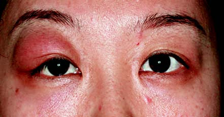
Figure 1 External appearance of acute dacryoadenitis in the right eye
Treatment
Treatment targets the underlying cause, involving systemic administration of antibiotics or antiviral medications in combination with topical application of antibiotic or antiviral eye drops to address bacterial or viral infections. If an abscess develops, timely incision and drainage are necessary. For eyelid lacrimal gland inflammation, drainage can be performed through a conjunctival incision, whereas abscesses in the orbital lacrimal gland can be drained via a skin incision.