Ectropion refers to an outward turning of the eyelid margin, causing it to separate from the eyeball. The palpebral conjunctiva is frequently exposed to varying degrees and is often accompanied by incomplete eyelid closure. Ectropion can be classified into degenerative ectropion, cicatricial ectropion, and paralytic ectropion, depending on the underlying cause.
Degenerative Ectropion
Degenerative ectropion, also referred to as senile ectropion, is limited to the lower eyelid. It occurs due to weakened orbicularis oculi muscle function and laxity of the eyelid skin and lateral canthal tendon in elderly individuals. These factors lead to the inability of the eyelid margin to maintain contact with the eyeball, and the downward displacement of the lower eyelid by its own weight contributes to the condition.
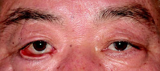
Figure 1 Appearance of degenerative ectropion
The patient shows tissue laxity and ectropion of the right lower eyelid.
Cicatricial Ectropion
Cicatricial ectropion results from scar contracture of the skin surface of the eyelid. It can be caused by trauma, burns, chemical injuries, eyelid ulceration, or previous eyelid surgeries.
Paralytic Ectropion
Paralytic ectropion is also confined to the lower eyelid and occurs as a result of facial nerve paralysis. The loss of orbicularis oculi muscle contraction causes the lower eyelid to turn outward due to its weight and loss of stability.
Clinical Manifestations
Mild ectropion is characterized by a slight outward turning of the eyelid margin, resulting in a loss of contact between the eyelid margin and the eyeball. This disrupts the siphon action between the eyelid and the eyeball, leading to epiphora (excessive tearing). Severe cases are characterized by exposed palpebral conjunctiva, which leads to the conjunctiva losing its protective moistening by tears. Initially, the exposed conjunctiva becomes congested and secretes excessive discharge; over time, it becomes dry, rough, and thickened, eventually developing a keratinized appearance. Additionally, ectropion is frequently associated with incomplete eyelid closure, leaving the cornea unprotected. As a result, the corneal epithelium may become dry and eroded, increasing the risk of exposure keratitis or corneal ulceration.
Diagnosis
Diagnosis is straightforward and based on the patient's medical history and characteristic clinical manifestations.
Treatment
Degenerative ectropion and cicatricial ectropion generally require surgical intervention. For paralytic ectropion, management focuses on addressing the underlying facial palsy. Additional measures include the use of eye ointment, mechanical traction of the eyelid to protect the conjunctiva and cornea, or temporary eyelid margin suturing.