Infectious blepharitis is commonly caused by bacterial, fungal, viral, or parasitic infections and typically presents as a chronic or subacute suppurative inflammation involving the eyelash follicles and their associated glands. The condition is often due to Staphylococcus aureus infection and is more frequently observed in children with malnutrition, anemia, or systemic chronic debilitating diseases.
Clinical Manifestations
Symptoms include severe itching, pricking sensations, and a burning sensation in the eyes. Small pustules are scattered around the bases of the eyelashes, covered with crusts that often cause the eyelashes to stick together in bundles. Removing the crusts reveals the bases of the eyelashes and shallow ulcers. In cases of Demodex mite infestation, "sleeve-like" changes around the eyelash roots may be visible. Eyelashes may fall out easily along with the crusts, and failure of the hair follicles to regenerate leads to areas of missing lashes (madarosis). Scar contraction following ulcer healing can alter the growth direction of eyelashes, resulting in misdirected growth (trichiasis) that can damage the cornea. Prolonged infection can lead to chronic conjunctivitis and thickening and deformation of the eyelid margin, eversion of the eyelid margin, and swelling or obstruction of the tear puncta, ultimately causing excessive tearing (epiphora).
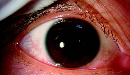
Figure 1 Appearance of infectious blepharitis
The patient exhibits "sleeve-like" changes around the bases of the eyelashes in the right upper eyelid due to Demodex mite infestation.
Diagnosis
The diagnosis relies on the typical clinical features, including the presence of pustules and ulcers on the eyelid margins.
Treatment
Performing bacterial cultures and antibiotic susceptibility testing is optimal for selecting effective treatments for infectious blepharitis.
Addressing Contributing Factors
Personal hygiene and removal of environmental triggers play a crucial role.
Eyelid Hygiene
The eyelid margins should be cleaned daily using normal saline or a 3% boric acid solution. This process removes crusts, detached eyelashes, and purulent discharge from the follicles. Massaging the eyelid margins with a cotton swab coated with antibiotic eye ointment can aid treatment.
Continuing Treatment After Recovery
Treatment should persist for at least 2–3 weeks after complete resolution of inflammation to minimize the risk of recurrence.
Anti-Mite Therapy
For cases involving Demodex mites, targeted anti-mite treatment is necessary.