Laryngeal keratosis refers to a keratotic and hyperplastic lesion of the epithelial lining of the laryngeal mucosa. It manifests as white, elevated patches on the mucosal surface, which are localized and not easily removed. The primary pathological changes involve varying degrees of thickening in the keratin layer of the squamous epithelium, which can include either hyperkeratosis or parakeratosis. The surrounding mucosa typically exhibits an inflammatory response, although the submucosal layer remains normal. This lesion can occur at any location within the laryngeal mucosa but is most commonly found on the vocal folds and in the interarytenoid region. Parakeratosis, when present, is specifically referred to as laryngeal leukoplakia.
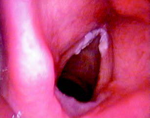
Figure 1 Laryngeal keratosis
Laryngeal keratosis and laryngeal leukoplakia may be associated with either simple epithelial hyperplasia or dysplasia to varying degrees. Those accompanied by epithelial dysplasia are regarded as precancerous lesions, with a malignant transformation rate of 3%–5%, and are more common in middle-aged men.
The exact etiology remains unclear. However, these conditions are generally linked to chronic inflammatory stimuli, such as smoking, alcohol consumption, vocal misuse, chronic laryngitis, reflux laryngopharyngitis, and deficiencies in vitamins or trace elements.
The main symptoms include hoarseness, throat irritation, coughing, and the sensation of a foreign body in the larynx.
Laryngoscopic examination often reveals chronic congestion of the laryngeal mucosa, with white conical lesions, plaques, or patches that are well-demarcated and firmly adhered to the mucosal surface, surrounded by a reddened, congested area. Dynamic (stroboscopic) laryngoscopy and narrow-band imaging (NBI) techniques can help in distinguishing precancerous lesions from early-stage laryngeal carcinoma.
Management depends on the degree of keratosis. Mild cases generally do not require specialized treatment. Avoidance of smoking, alcohol consumption, and other factors that might irritate the laryngeal mucosa is recommended. Long-term use of vitamin A supplements may have some therapeutic effects.
For cases with more pronounced keratosis, surgical removal of the lesion under a suspension laryngoscope is often performed. Techniques such as vocal fold stripping or CO2 laser surgery may be applied. For extensive or rapidly progressing lesions, laryngofissure combined with vocal fold stripping surgery may be used.
Laryngeal keratosis or leukoplakia with epithelial dysplasia, due to its classification as a precancerous condition, requires close observation and regular follow-up. Patients with severe dysplasia or carcinoma in situ should undergo surgery at the earliest opportunity.