The maxilla is connected to several bones, including the frontal bone, zygomatic bone, nasal bone, vomer, ethmoid bone, lacrimal bone, sphenoid bone, and palatine bone. Fractures of the maxillary sinus often form part of a complex maxillofacial fracture with associated features. Midface fractures primarily involve fractures of the maxilla, affecting the midsection of the craniofacial skeleton. These fractures can span multiple craniofacial bones and are typically open fractures. The most common cause is motor vehicle accidents, and the injuries are often complex and severe, sometimes requiring the cooperation of neurosurgery, maxillofacial surgery, and ophthalmology for treatment. Due to the presence of sinus cavities, spaces, and joints inside and around the maxilla, the injury often communicates with the oral cavity, nasal cavity, and maxillary sinus, thus raising the risk of infection.
Clinical Manifestations
Manifestations of Maxillary Fractures
Fractures of the maxilla are most often caused by direct impact trauma. Different areas of involvement lead to varying presentations, including localized swelling, deformity with collapse, and asymmetry of the maxillofacial region. After the swelling subsides, the deformity becomes more pronounced, affecting facial aesthetics and functional movements. Fractures involving the nasal cavity mucosa and nasal sinus mucosa may result in nasal bleeding. Fractures of the nasal bone or displacement of the nasal septum bone produce corresponding symptoms. Compression of the infraorbital nerve due to fractures involving the infraorbital foramen can cause numbness in the infraorbital region and upper lip. Maxillary sinus roof fractures or fractures involving the orbital floor may lead to enophthalmos, impaired ocular movement, and diplopia. When fracture lines extend to the frontal sinus, ethmoidal sinus, or sphenoid sinus, cerebrospinal fluid rhinorrhea may occur; fractures involving the petrous part of the temporal bone may result in cerebrospinal fluid otorrhea. Displacement of maxillary fractures involving the zygomatic bone may lead to restricted mouth opening or malocclusion.
Classification of Midface Fractures
Horizontal Fractures (Le Fort type I)
These fractures occur due to a downward force acting on the lower portion of the maxilla. The fracture line extends from the base of the piriform aperture, traversing the root of the alveolar process, the partial walls of the maxillary sinus, the base of the nasal septum, and the maxillary tuberosity, continuing horizontally posteriorly through the pyramidal process of the palatine bone to the lower portions of the pterygoid plates of the sphenoid bone. This type causes a complete fracture of the structures beneath the alveolar process, palatine bone, and maxillary tuberosity. Typical features include downward displacement of the upper alveolar arch and nasal cartilage, swelling of the upper lip, loosening of upper teeth, and elongation of the facial appearance. Hematoma or bleeding may occur in the gingival sulcus, hard palate, or anterior-inferior nasal septum. Movement of the upper teeth may mobilize the entire fractured segment.
Pyramidal Fractures (Le Fort type II)
These result from impacts to the middle portion of the maxilla. The fracture line traverses the nasal bone, nasofrontal suture, nasal septum, and frontal process of the maxilla. It then extends bilaterally through the lacrimal bone, infraorbital margin, and obliquely downward to the orbital floor, passing posteriorly through the lateral wall of the maxillary sinus, pterygoid plates, and pterygopalatine fossa. Severe cases may cause lateral displacement of the ethmoidal and lacrimal bones, widening the interorbital distance. Separation of the zygomatic-maxillary complex may cause the inferior zygomatic bone to sink. Clinical features include marked facial swelling, subconjunctival hemorrhage, and bruising and swelling of both eyelids, creating a bluish discoloration. Posterior and downward displacement of fractured segments causes midfacial flattening and facial elongation, giving a "dish-face" appearance. Movement of the upper facial segment during jaw opening and closing may be observed. Posterior displacement of fractures and soft palate swelling may lead to obstructive airway issues.
Craniofacial Separation Fractures (Le Fort type III)
This fracture occurs when force impacts the uppermost weak line of the maxillary body. The fracture line originates from the nasal bone and nasofrontal suture, extends laterally through the frontal process, then descends along the medial orbital wall and orbital floor to the inferior orbital fissure. It traverses the zygomaticofrontal suture above the zygomatic bone and extends posteriorly to the root of the pterygoid process of the sphenoid bone. This fracture completely separates the middle third of the face from the cranial base, leaving the midfacial bones connected solely by soft tissues.
This injury is severe and complex, often accompanied by cranial brain trauma, basal skull fracture, orbital injuries, ocular damage, or optic nerve injury. Clinical manifestations may include coma, neck stiffness, paralysis of related cranial nerves, sudden vision loss, intraorbital hematoma/emphysema, exophthalmos, and inferior displacement of the eyeball.
Others
The classical Le Fort classification of fractures is not commonly observed in clinical practice. Most cases involve mixed or combined fractures. Fracture lines on both sides are often not on the same plane or do not belong to the same type, and unilateral fractures may also occur. Therefore, Marciani proposed a modified classification method for maxillary fractures in 1993:
- Le Fort Type I refers to horizonal maxillary fractures, with Type Ia indicating horizontal multiple fractures of the maxilla;
- Le Fort Type II refers to pyramidal maxillary fractures, with Type IIa involving concurrent nasal bone fractures and Type IIb involving concurrent naso-orbital-ethmoid fractures;
- Le Fort Type III refers to craniofacial maxillary fractures, with Type IIIa involving concurrent nasal bone fractures and Type IIIb involving concurrent naso-orbital-ethmoid fractures;
- Le Fort Type IV refers to Le Fort Type II or III fractures combined with skull base fractures, with Type IVa involving supraorbital margin fractures, Type IVb involving supraorbital margin and anterior cranial fossa fractures, and Type IVc involving orbital wall and anterior cranial fossa fractures.
Diagnosis
Based on the history of trauma, clinical presentation, craniofacial deformity, palpation revealing a depression, and numbness in the infraorbital region and upper lip, the diagnosis can be confirmed. Maxillofacial CT imaging can identify the fracture site, while 3D reconstruction provides a clear visualization of the stereoscopic anatomical relationships. It is important not to overlook severe cranial brain injuries, optic nerve injuries, and other serious complications such as cerebrospinal fluid rhinorrhea.
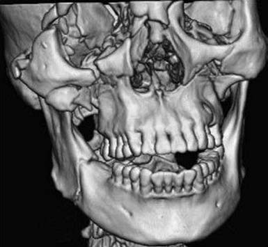
Figure 1 Three-dimensional reconstruction image of a maxillary fracture from a CT scan
Treatment
Emergency care needs prompt management. The treatment principles focus on controlling bleeding and maintaining airway patency, with tracheotomy performed when necessary. Once vital signs stabilize, fracture reduction and fixation are carried out, involving collaboration with relevant departments. For depressed fractures of the anterior wall of the maxillary sinus, reduction is performed through the intraoral upper gingivobuccal sulcus approach, followed by internal fixation using micro titanium plates. Fractures of the superior wall of the maxillary sinus, including the orbital floor, are addressed through approaches such as the subciliary incision or transconjunctival incision, utilizing artificial materials or autologous bone for reconstruction. Precise and accurate reduction of the zygomatic bone, orbit, maxilla, and orbital floor is critical for early-stage fracture repair surgery.