Tumors of the external ear can arise from various tissues in the auricle and external auditory canal, including skin, blood vessels, nerves, and bony structures. Common pathological types of tumors in this area include basal cell carcinoma, exostoses, papillomas, ceruminous gland tumors, and hemangiomas. Most of these tumors are primary and benign, while a small proportion are malignant. Malignant tumors often result from the malignant transformation of primary benign tumors.
Benign Tumors
Papillomas of the Auricle or External Auditory Canal
Papillomas, one of the most common benign tumors of the ear, often involve the skin surface of the auricle or the cartilaginous portion of the external auditory canal. These tumors are more common in males and are generally believed to be associated with viral infections or chronic local irritation. Approximately 2% of cases show a potential for malignant transformation.
Clinical Symptoms and Signs
Early stages are often asymptomatic. When the tumor fills the external auditory canal, symptoms may include a sense of obstruction, hearing loss, or itching. Secondary infection may lead to discharge, otalgia, or pain. Physical examination typically reveals single or multiple uneven-surfaced tumors of varying sizes within the external auditory canal. The lesions are usually firm on palpation and often have broad bases. In rare cases, extension into the middle ear may occur.
Diagnosis and Treatment
Diagnosis can be made based on medical history and otological examination. Definitive diagnosis requires pathological biopsy, which generally shows papillary structures with a fibrous vascular core covered by squamous epithelium that protrudes towards the surface. The upper layers of cells often display incomplete or excessive keratinization. Differential diagnosis should include external auditory canal cancer and verruca vulgaris.
Treatment primarily involves surgical excision. Incomplete removal increases the risk of recurrence. Postoperative application of silver nitrate, Brucea javanica oil, or interferon on the wound may help reduce the risk of recurrence. Malignant potential increases, especially when the middle ear is involved, necessitating close postoperative monitoring.
Exostoses of the External Auditory Canal
Exostoses of the external auditory canal are nodular protuberances resulting from localized overgrowth of bony tissue in the bony walls of the external auditory canal. These are more common in adult males, typically bilateral and multiple. The exact etiology is uncertain, although local trauma, inflammation, and exposure to cold water are potential contributing factors.
Clinical Symptoms and Signs
Small exostoses are frequently asymptomatic and may only be discovered incidentally during routine otological examinations or cerumen removal. Larger lesions may cause narrowing of the external auditory canal and lead to secondary conditions such as cholesteatoma formation, often accompanied by hearing loss. Infections may cause symptoms such as otorrhea, otalgia, or bleeding from the ear.
Diagnosis and Treatment
Smooth, hard nodules located on the bony portion of the external auditory canal suggest the possibility of exostoses. Solitary exostoses may be misdiagnosed as cysts or polyps, but these can be distinguished by their softer consistency.
Treatment is not required for asymptomatic lesions. For symptomatic cases or those causing obstruction of the external auditory canal, early surgical resection is recommended.
Osteomas of the External Auditory Canal
Osteomas are rare benign tumors of the external auditory canal. They are typically unilateral and can lead to complications such as conductive hearing loss and external auditory canal inflammation. These tumors are solitary, bony, slow-growing, and hard, with surfaces generally covered by normal skin. The etiology is unclear, but possible contributing factors include chronic irritation, trauma, or infection leading to abnormal bone growth.
Clinical Symptoms and Signs
Symptoms tend to appear relatively early because these lesions grow more rapidly than exostoses and often have a broad base. Symptoms may include external auditory canal obstruction with subsequent narrowing, secondary cholesteatoma, hearing loss, and occasionally otorrhea, otalgia, or ear bleeding.
Diagnosis and Treatment
Diagnosis is typically straightforward and based on medical history and otological examination. Temporal bone CT can provide detailed information on the tumor's size, location, and involvement of structures such as the mastoid or tympanic cavity. Surgical excision is the treatment of choice.
Ceruminous Gland Tumors of the External Auditory Canal
These rare tumors originate from the epithelial and myoepithelial cells of ceruminous gland ducts within the cartilaginous portion of the external auditory canal. They are most frequently found in the floor and anterior wall of the external auditory canal and are characterized by slow growth.
Clinical Symptoms and Signs
In the early stages, tumors are often asymptomatic. As they enlarge, they may lead to non-specific symptoms such as a sensation of ear blockage, ear pain, and hearing impairment. Secondary infections may result in discharge mixed with blood, exacerbated ear pain, and other manifestations. Ceruminous gland tumors typically appear as grayish-white, polypoid masses with smooth surfaces covered by normal skin and are firm in texture.
Diagnosis and Treatment
Definitive diagnosis is established through pathological examination. Specific situations that warrant consideration of ceruminous gland tumors in the external auditory canal include:
Granulation tissue in the external auditory canal does not resolve with standard treatments.
Swelling and protrusion of the external auditory canal wall, accompanied by bloody discharge.
Persistent localized pain in the external auditory canal unresponsive to conventional therapies.
These tumors are insensitive to radiation therapy, and surgical excision is the preferred treatment. While ceruminous gland tumors and pleomorphic adenomas are histologically benign, both have high recurrence and malignant transformation rates. As such, clinical management follows principles for treating tumors with malignant tendencies.
Other Common Benign Tumors of the Auricle and External Auditory Canal
Hemangiomas
Hemangiomas are common in the auricle and may extend to the periauricular skin or skin of the external auditory canal. Capillary hemangiomas and cavernous hemangiomas are most frequently observed, while compacted hemangiomas and arterial hemangiomas are less common. All are benign tumors, and management options include surgical excision, laser therapy, cryotherapy, or sclerosant injections.
Cysts
Cysts can occur on the auricle or periauricular area and are most commonly found on the auricle. Examples include sebaceous cysts and epidermoid cysts, with cyst walls lined by epithelium. These structures develop slowly and require complete excision for treatment.
Fibromas
Fibromas predominantly occur on the auricle and are classified into soft and hard types based on the fibrous and cellular composition of the tumor tissue. Soft fibromas contain fewer fibers and abundant tumor cells, resembling lipomas, while hard fibromas mostly consist of collagen fibers and present as firm, painless nodules. Surgical excision is the standard treatment for both types.
Malignant Tumors
Most malignant tumors of the external ear originate from the skin, with squamous cell carcinoma being the most prevalent, accounting for approximately 60% of all external ear malignancies. Basal cell carcinoma follows as the second most common type. The development of squamous cell carcinoma in the external ear is generally associated with prolonged sun exposure, chronic infections, and recurrent irritation.
Squamous Cell Carcinoma of the External Auditory Canal
Clinical Symptoms
Typical symptoms include otalgia and bloody discharge. Secondary infections may lead to purulent otorrhea and hearing loss. When the facial nerve is involved, peripheral facial paralysis may occur. This condition is most common among middle-aged and elderly men and is often misdiagnosed as chronic inflammation during the early stages, leading to delayed treatment. Biopsy is recommended if symptoms persist despite appropriate treatment. Tumor staging follows the Pittsburgh staging system (TNM classification).
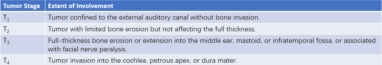
Table 1 Pittsburgh staging system for external auditory canal tumors
Note: Lymph node staging (N) and distant metastasis staging (M) follow the same criteria as those used for other head and neck tumors.
Treatment
Management includes surgical excision followed by adjuvant radiotherapy. For T1 and T2 lesions, lateral temporal bone resection is performed, along with superficial parotidectomy and selective neck dissection (levels II–III). For T3 and T4 lesions, subtotal or total temporal bone resection is required.
Adenoid Cystic Carcinoma
Adenoid cystic carcinoma is a low-grade malignancy characterized by slow growth and a propensity for perineural invasion. It frequently metastasizes to the lungs, but distant metastases are not considered contraindications for local surgery.
Clinical Symptoms
Symptoms include ear pain, a mass in the external auditory canal, hearing loss, tinnitus, and discharge, which are non-specific overall. The tumor may be poorly demarcated from adjacent tissues, firm in texture, and tender upon palpation. It can also present as polyps, ulcers, or granulation tissue. Diagnosis requires pathological biopsy.
Treatment
Surgical excision remains the primary treatment. Depending on the extent of invasion, procedures may involve concomitant superficial or total parotidectomy, neck dissection, and lateral temporal bone resection. Postoperative radiotherapy is generally less effective.
Ceruminous Adenocarcinoma
Clinical Symptoms
Ceruminous adenocarcinoma typically manifests as painless external auditory canal bleeding or susceptibility to bleeding when cleaning the ear. Ear pain may occasionally occur. Tumors generally appear as reddish, granular masses with irregular, rough surfaces. When the tumor invades the parotid gland by breaking through the cartilaginous part of the external auditory canal, parotid swelling may be observed. Forward invasion into the temporomandibular joint may result in difficulty opening the mouth.
Treatment
Surgical excision is the primary approach, supplemented by radiotherapy, although recurrence is common. Postoperative radiotherapy reduces the likelihood of tumor recurrence.
Malignant Melanoma
Clinical Presentation
Malignant transformation into melanoma should be considered when pigmented nevi, following mechanical irritation (such as repeated friction or scratching), present with ulceration, bleeding, or pain, accompanied by rapid growth of the mass or the appearance of satellite lesions.
Treatment
Complete surgical excision is the main treatment. Preoperative biopsy is generally avoided to prevent accelerated tumor growth and metastasis. For extensive tumors, wide excision of the external auditory canal, mastoidectomy, and, if necessary, procedures such as parotidectomy, subtotal temporal bone resection, or cervical lymph node dissection are performed, though the prognosis remains poor.
Melanoma, known for its immunogenic characteristics, has been shown to respond poorly to chemotherapy. Significant progress has been made in understanding the molecular mechanisms and signaling pathways involved in its development, and immunotherapy has increasingly been applied in clinical settings.