Acute suppurative otitis media (ASOM) is an acute purulent inflammation of the middle ear mucosa. It is more common in children, frequently occurs in the winter and spring seasons, and is often secondary to upper respiratory tract infections.
Etiology
The main causative pathogens include Streptococcus pneumoniae, Haemophilus influenzae, Streptococcus pyogenes, and Staphylococcus aureus. Common routes of infection include:
- Eustachian tube route
- External auditory canal and tympanic membrane route
- Hematogenous spread
Eustachian Tube Route
Bacteria invade the middle ear via the Eustachian tube, leading to acute upper respiratory tract infections.
Acute infectious diseases such as scarlet fever, measles, and pertussis can cause this condition as a complication via the Eustachian tube. It may also manifest as a localized symptom of these infections.
Activities such as swimming, diving, improper nose-blowing techniques, Valsalva maneuvers, and nasal treatments can allow bacteria to enter the middle ear through the Eustachian tube.
The shorter, wider, and more horizontally positioned Eustachian tube in infants facilitates the retrograde entry of bacteria or secretions from the nasopharynx into the middle ear. In case of supine feeding, milk or gastric contents may reflux into the middle ear via the Eustachian tube.
External Auditory Canal and Tympanic Membrane Route
Direct bacterial entry into the middle ear can be through tympanic membrane perforation, tympanostomy tube placement, or tympanic membrane trauma.
Hematogenous Spread
Rarely, the infection can spread to the middle ear through the bloodstream.
Pathology
In the early stages of infection, the middle ear mucosa becomes congested and edematous, with closure of the Eustachian tube's nasopharyngeal orifice. Air in the tympanic cavity is resorbed, creating negative pressure. Inflammatory exudates gradually become purulent. As pus accumulates, pressure in the tympanic cavity increases, leading to localized bulging of the tympanic membrane. Thrombotic venulitis can cause necrosis and rupture of the tympanic membrane, resulting in perforation and pus drainage. Small tympanic membrane perforations may heal spontaneously.
Clinical Manifestations
Before tympanic membrane perforation, most patients experience severe pain, often described as pulsating or stabbing pain, which may radiate to the ipsilateral head or teeth. After tympanic membrane perforation and pus drainage, ear pain typically subsides.
In the early stages, patients often report a sense of ear fullness, low-pitched tinnitus, and hearing loss. After tympanic membrane perforation and pus drainage, hearing loss may improve.
In patients with severe ear pain, hearing impairment may be overlooked. A minority of patients may experience vertigo.
Pus drainage occurs after tympanic membrane perforation. Initially, the discharge is purulent and blood-tinged, later becoming mucopurulent.
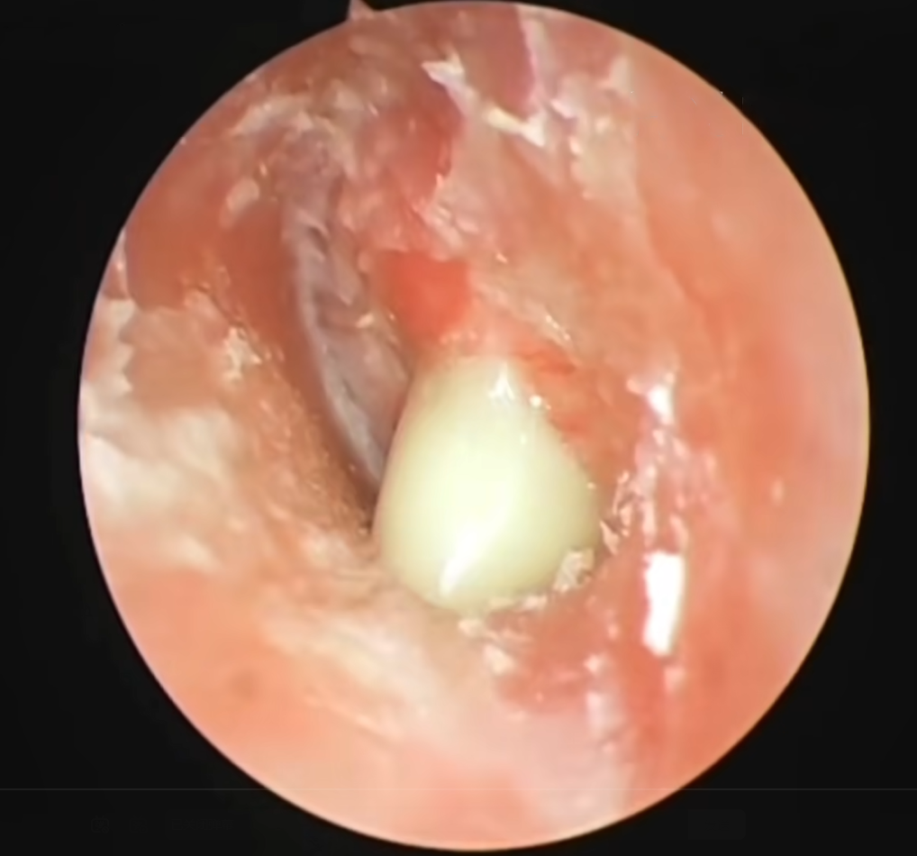
Figure 1 Acute suppurative otitis media
Patients may experience chills, fever, fatigue, and anorexia.
In children, systemic symptoms are often more severe and may include emesis, diarrhea, and other symptoms resembling gastrointestinal infection. Once the tympanic membrane perforates, body temperature quickly normalizes, and systemic symptoms significantly improve.
In the early stages, the pars flaccida of the tympanic membrane shows congestion, with radiating dilated blood vessels visible around the handle of the malleus and the tense portion of the tympanic membrane.
As the condition progresses, the tympanic membrane becomes diffusely congested, swollen, and outwardly bulging, with normal landmarks disappearing. Small yellow spots may appear, which can progress to tympanic membrane perforation.
Initially, perforations are small and may be difficult to detect. Pus may flow out from the perforation, forming a pulsating bright spot known as lighthouse sign. In necrotizing cases, the tympanic membrane may rapidly disintegrate, resulting in large perforations.
Mild tenderness may be present over the mastoid area, with more pronounced tenderness in the mastoid antrum region.
Hearing loss is primarily conductive in most patients. In rare cases, cochlear involvement may lead to mixed or sensorineural hearing loss.
An increase in total white blood cell count and neutrophils is commonly observed. After tympanic membrane perforation, blood test results gradually return to normal.
Diagnosis
The diagnosis can be established based on the patient’s medical history and clinical manifestations.
Differential Diagnosis
Acute Otitis Externa and Furuncle of the External Auditory Canal
They are characterized by significant ear pain and tenderness when the auricle is pulled. Swelling is observed at the external auditory canal opening and within the canal, which may progress to a localized furuncle in the late stage. The tympanic membrane shows mild or no inflammation, and hearing is generally normal.
Acute Myringitis
It is often associated with influenza or herpes zoster oticus. Severe ear pain is present, but hearing loss is usually not significant. Examination reveals tympanic membrane congestion with bullae. Tympanic membrane perforation is typically absent.
Prevention
Proper techniques for nose-blowing and breastfeeding are required. Upper respiratory tract infections and respiratory infectious diseases should be properly treated. For patients with tympanic membrane perforation or tympanostomy tubes, water entering the ear should be avoided.
Treatment
Treatment principles include controlling infection, ensuring proper drainage, and addressing the underlying cause.
Systemic Treatment
Adequate doses of antibiotics should be administered early to control the infection. Commonly used antibiotics include penicillins and cephalosporins.
For patients with tympanic membrane perforation, pus should be collected for bacterial culture and drug sensitivity testing to guide antibiotic selection.
For severe systemic symptoms, supportive therapies such as fluid replacement can be provided.
Local Treatment
Before Tympanic Membrane Perforation
1% phenol-glycerin ear drops can be used to reduce inflammation and relieve pain. Nasal drops containing vasoconstrictors can be applied to improve Eustachian tube patency and reduce local inflammation. If systemic and local symptoms are severe, and the tympanic membrane is significantly bulging without improvement after standard treatment, myringotomy can be performed under sterile conditions.
For redness, swelling, and tenderness in the posterosuperior part of the auricle, acute mastoiditis may be suspected. If confirmed by CT scan, mastoidectomy with drainage can be considered.
After Tympanic Membrane Perforation
3% hydrogen peroxide solution can be used to clean and remove pus from the external auditory canal, or suction can be used to remove it.
Antibiotic solutions can be used as ear drops. Powders should be contraindicated to prevent clumping with pus, which can obstruct drainage.
When pus decreases and inflammation subsides, alcohol-based ear drops with a dehydrating effect, such as 3% boric acid ethanol-glycerin or 3% boric acid ethanol, may be used as appropriate.
Chronic conditions of the nasal cavity, sinuses, pharynx, and nasopharynx should be properly treated to prevent recurrence of otitis media. After infection is completely controlled and inflammation is resolved, tympanic membrane perforation may heal spontaneously in some patients.