External auditory canal cholesteatoma, also known as obstructive keratosis of the external auditory canal, is a mass of desquamated epithelial debris containing cholesterol crystals that obstructs the bony portion of the external auditory canal.
Etiology
The condition is thought to be associated with chronic irritation of the external auditory canal skin caused by factors such as cerumen impaction, inflammation, foreign bodies, fungal infections, and other stimuli. These irritants may lead to chronic congestion, causing the basal cells in the germinal layer of the local skin to grow actively. This results in an abnormal increase in the shedding of keratinized epithelial cells. If the discharge of these cells is obstructed, they accumulate in the external auditory canal, forming a mass. Over time, the center of the mass undergoes necrosis, decomposition, and degeneration, leading to the formation of cholesterol crystals and the development of a cholesteatoma.
Clinical Manifestations
This condition occurs mostly in adults and is usually unilateral, though it can affect both ears.
A large cholesteatoma may cause symptoms such as a sense of blockage in the ear, tinnitus, and hearing loss.
If secondary infection occurs, symptoms may include ear pain, headache, and foul-smelling discharge from the external auditory canal.
Examination reveals white or yellow cholesteatoma obstructing the deep portion of the external auditory canal, with its surface covered by multiple layers of scaly debris.
Removal of a large cholesteatoma may reveal bony destruction and resorption of the external auditory canal, with significant widening of the bony portion.
A massive cholesteatoma can destroy the posterior wall of the external auditory canal, invade the middle ear, extensively damage the mastoid bone, and lead to complications such as cholesteatomatous otomastoiditis. It may also cause peripheral facial paralysis.
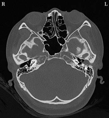
Figure 1 The cholesteatoma in the left external auditory canal damaging the posterior wall of the external auditory canal
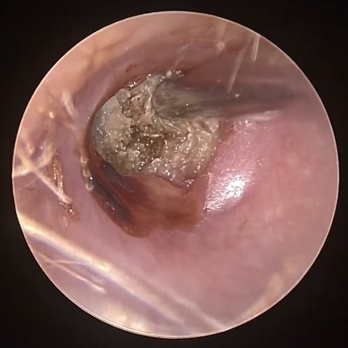
Figure 2 Cholesteatoma in the external auditory canal
Diagnosis
Diagnosis is based on the patient’s history and the characteristic white cholesteatoma mass in the external auditory canal. Pathological examination can confirm the diagnosis. It is important to differentiate external auditory canal cholesteatoma from cholesteatoma originating in the middle ear, external auditory canal carcinoma, and necrotizing external otitis. Temporal bone CT scans or mastoid x-rays may be performed if necessary.
Treatment
For cholesteatomas without infection, they are relatively easy to remove and can be extracted using a cerumen hook.
For cholesteatomas with secondary infection, controlling the infection is essential. However, infection control alone is often insufficient for rapid improvement. Partial or complete removal of the cholesteatoma is necessary to facilitate the resolution of inflammation.
For severe infections or cases where removal is difficult, surgery may be performed under general anesthesia and with the aid of a surgical microscope. Systemic antibiotics should be administered to control the infection. Postoperative follow-up is required to monitor for and remove any residual or recurrent cholesteatoma.
For extensive disease, otoplasty may be necessary. In some cases, mastoidectomy may also be required.