Suppurative perichondritis of the auricle is an acute purulent inflammation of the perichondrium of the auricle, occurring between the cartilage and perichondrium. It often causes severe pain and can lead to cartilage necrosis and auricular deformity if not promptly and properly treated.
Etiology
The condition is commonly caused by secondary infection following trauma, surgery, frostbite, burns, or auricular hematoma. The most frequently isolated pathogen is Pseudomonas aeruginosa, followed by Staphylococcus aureus.
Clinical Manifestations
The initial symptoms include swelling and pain of the auricle, which progressively worsen with increasing redness, swelling, fever, and pain over an expanding area. Patients may experience significant discomfort, accompanied by systemic symptoms such as fever and anorexia. On examination, the auricle appears red, swollen, and tender. Once an abscess forms, undulation may be present, and the abscess may rupture, discharging pus.
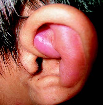
Figure 1 Suppurative perichondritis of the auricle
Treatment
When an abscess has not yet formed, systemic administration of sensitive antibiotics is essential to control the infection. Local physical therapy, such as heat application, may also be employed to promote resolution of the inflammation.
If an abscess has formed, surgical drainage is necessary under general or local anesthesia. A semicircular incision is made along the scaphoid fossa on the medial side of the helix to fully expose the abscess cavity. Pus is thoroughly drained, granulation tissue is removed, and necrotic cartilage is excised. Preserving the cartilage in the helix region, if possible, can prevent auricular deformity. The surgical cavity should be thoroughly irrigated. After surgery, the skin is repositioned over the wound, and a skin flap drain is placed without complete closure of the incision to prevent postoperative hematoma or organization and contraction. Appropriate compression bandaging is applied, and dressings are changed every other day or daily. Alternatively, a small tube with fine perforations can be placed in the surgical cavity, and the wound is sutured in alignment. The cavity is irrigated daily until both local and systemic symptoms resolve, after which the tube is removed.
For patients with severe auricular deformities, reconstructive surgery may be performed.
Prevention
During procedures involving the auricle, such as surgery or ear piercing, strict aseptic techniques should be followed and cartilage injury should be avoided. Auricular trauma should be promptly treated with thorough debridement to prevent infection.