An aural pseudocyst is a non-suppurative serous fluid accumulation within the cartilage layers of the auricle, forming a pseudocyst. It typically occurs on the upper half of the lateral anterior surface of one auricle and contains serous exudate, presenting cystic swelling. This condition is also known as non-suppurative perichondritis of the auricle, serous perichondritis of the auricle, or intracartilage effusion of the auricle. It commonly affects one auricle, is more common in males than females, and typically occurs in adults aged 20 - 50.
Etiology
The exact cause is unclear but may be related to trauma, mechanical irritation, or congenital developmental factors.
Pathology
The cyst is located between the cartilage layers in microscopy. Therefore, from a pathological perspective, it is more accurately described as an intracartilage effusion. The inner surface of the cartilage layer is covered by a serofibrinous layer without epithelial cell structures, which distinguishes it from a true cyst.
Clinical Manifestations
The cystic swelling is usually located in the scaphoid fossa or triangular fossa, and occasionally extends to the conchal cavity, but does not affect the posterior surface of the auricle. Patients often notice a localized swelling on the upper anterior part of the auricle, which gradually enlarges. Small cysts may be asymptomatic, while large ones may cause sensations of fullness, undulation, burning, or pruritus, though they are often painless or only mildly tender. The cyst has a well-defined border, and the overlying skin appears normal in color. When transilluminated, the cyst exhibits good translucency, which helps differentiate it from a hematoma. Aspiration yields clear, pale yellow fluid, and bacterial cultures show no growth.
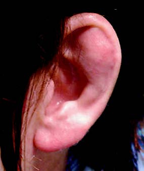
Figure 1 Aural pseudocyst
Treatment
Physical Therapy
In the early stages, physical therapies such as ultraviolet irradiation or ultrashortwave treatment can be performed to reduce exudation and promote resorption. Other reported treatments include wax therapy, magnetic therapy, cryotherapy, and radiofrequency therapy.
Aspiration and Local Compression
Under strict aseptic conditions, the cyst fluid can be aspirated, followed by local compression using plaster fixation or pressure with fine gauze strips secured with bandages. Alternatively, two round magnets (approximately 1.5 cm in diameter) can be placed on the anterior and posterior surfaces of the auricle at the site of the cyst, using magnetic force to apply compression.
Intracystic Injection of Medications
Some reports suggest injecting medications such as bleomycin, 15% hypertonic saline, and 50% glucose solution into the cyst cavity after aspiration. No compression is applied afterward. The injected fluid is aspirated in 24 hours, and the procedure is repeated until the aspirated fluid becomes reddish, promoting adhesion and organization of the cyst wall. However, the cure rate is suboptimal, and the treated area often becomes thickened or deformed.
Surgical Treatment
Surgery generally yields favorable outcomes in most cases. The procedure involves incising the outer cartilage wall of the cyst, completely aspirating the fluid, and removing any granulation tissue if present. A drainage strip can be placed in the surgical cavity, and the incision is sutured and compressed with a dressing for approximately two days.